
Presentation
History of hand trauma
Patient Data
Age: 35 years
Gender: Male
From the case:
Triangular fibrocartilage complex displaced tear


Download
Info
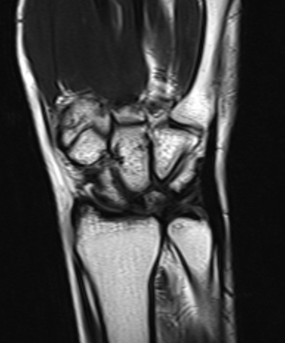
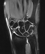
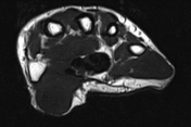
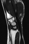
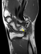
Multiple MRI sections of the wrist joint show a full-thickness tear within the central triangular fibrocartilage that displaced toward the volar aspect of the distal radioulnar joint and was associated with fluid accumulation around this region. These features are consistent with a displaced tear of the triangular fibrocartilage complex.
From the case:
Triangular fibrocartilage complex displaced tear
Download
Info
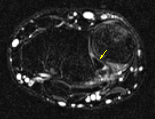
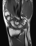
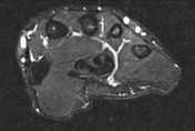
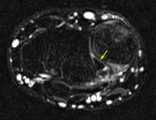
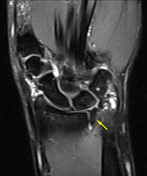
Selected MRI images clarified the displaced component of the triangular fibrocartilage complex (yellow arrow).
Case Discussion
The triangular fibrocartilage complex is a complicated structure and needs careful evaluation by MRI imaging.


 Unable to process the form. Check for errors and try again.