
Presentation
Optic neuritis ?
Patient Data
Age: 55 years
Gender: Female
From the case:
Tuberculum sellae meningioma


Download
Info
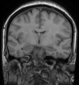
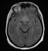
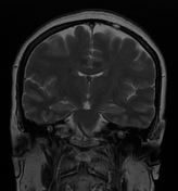
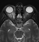
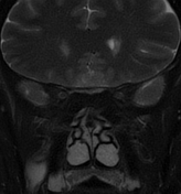
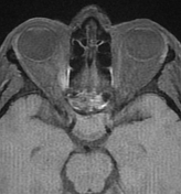
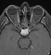
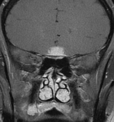
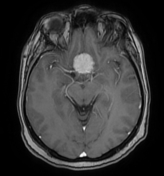
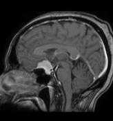
There is a well-defined sellar/suprasellar mass with a suprasellar epicentre. It displays an isosignal to the cortical grey matter on both T1/T2 with a vivid homogeneous enhancement on postcontrast sequences with a dural tail sign. The suprasellar component compresses the optic chiasma and the intracranial segment of the optic nerves which are displaced laterally.
Homogeneous enhancement of the pituitary gland, well-visualised on all sequences. The pituitary stalk is displaced posteriorly.
Case Discussion
MRI features most consistent with a tuberculum sellae meningioma.


 Unable to process the form. Check for errors and try again.