
Presentation
Visual disturbances.
Patient Data
Age: 40 years
Gender: Female
From the case:
Tuberculum sellae meningioma

Show annotations
Download
Info
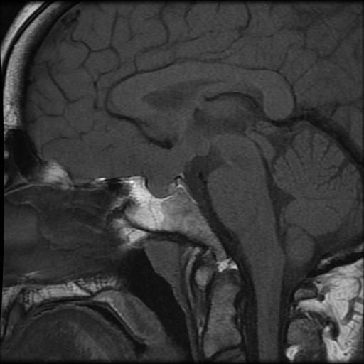
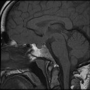
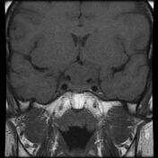
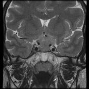
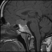
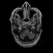
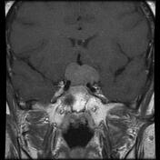
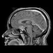
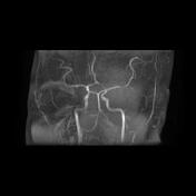
Well-defined lobulated extra-axial sellar and suprasellar mass centred on the tuberculum sellae. It displays an isosignal to the cortical grey matter on T1 and T2 with a homogeneous enhancement on postcontrast sequences. The suprasellar component compresses the optic chiasma and encases partially the supraclinoid segment of both ICA (mainly on the left) which show reduced calibre on the MRA.
The pituitary gland is well-visualised on enhanced sagittal and coronal T1 sequences.
Case Discussion
MRI features most consistent with a tuberculum sellae meningioma compressing the optic chiasma.


 Unable to process the form. Check for errors and try again.