
Presentation
Seizures since 1 month.
Patient Data
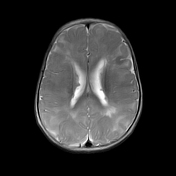

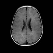
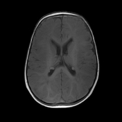
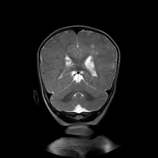
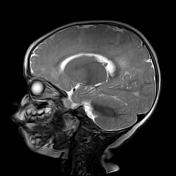
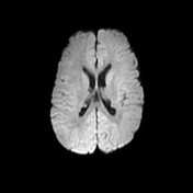
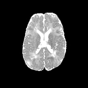
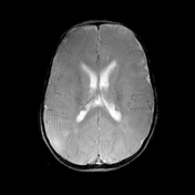

Multiple subependymal nodules are present in both the lateral ventricles which appear isointense to brain parenchyma on all imaging sequences. No evidence of calcification in these subependymal nodules.
Multiple T2 hyperintensities are seen in cortical and subcortical white matter of both cerebral hemispheres - likely represent cortical tubers.
Case Discussion
Cortical and subcortiucal tubers are seen in most of the cases and occurs due to enlarged atypical and disoragnized neuronal and glial elements with astrocytosis. Cortical tubers are typically benign pathologic entities. However, the extent of cerebral dysfunction, including intractable epilepsy and mental retardation, may be related to the burden from cortical tubers, especially when there is bilateral hemisphere involvement. In above case it acted as epileptogenic focus.
Subependymal nodules are similar to cortical tubers but have a tendency to calcify with age. It may convert into subependymal giant cell astrocytomas and occur near foramen of monro. It may result in obstructive hydrocephalus and patient requires survelliance every 1-3 years to rule out SEGA's.


 Unable to process the form. Check for errors and try again.