
Presentation
The patient presented with seizures.
Patient Data
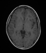

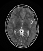
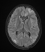
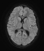
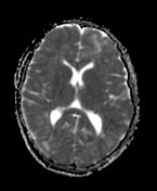
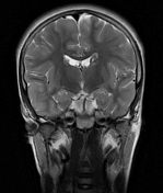
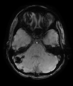
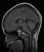
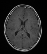
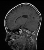

The MRI sequences demonstrate:
extensive bilateral cortical and subcortical areas of abnormal signal intensity with low/isointense signal on T1, high signal on T2 and more obvious with high signal intensity on FLAIR
bilateral subependymal nodules with blooming artifact on SWI suggesting calcified nodules and reveal no appreciable enhancement after IV contrast stud
bilateral radial bands of high signal on T2 and FLAIR extending from the periventricular white matter to the subcortical regions and fanning out as they reach the periphery
large right cerebellar contiguous wedge shaped lesions with blooming artifact, representing cerebellar tuber
Case Discussion
The radiological findings of bilateral subcortical tubers, subependymal calcified nodules, radial band signs and cerebellar wedge shaped calcified tuber are typical and characteristic of tuberous sclerosis.


 Unable to process the form. Check for errors and try again.