
Presentation
History of head trauma three years back followed by fits for one year.
Patient Data

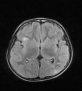
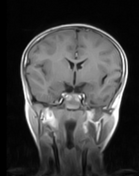
Multiple small subependymal nodules without definite enhancement involving walls of the bilateral lateral ventricles are noted.
Multiple areas of variable-sized altered signal intensity are noted in the bilateral cerebral hemispheres involving subcortical and cortical regions. The returning signals are hypointense on T1WI, hyperintense on T2WI and hyperintense on FLAIR sequence.
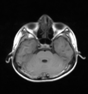
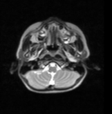
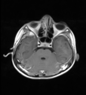
Patchy enhancement is seen in the bilateral cerebelli.
Case Discussion
MRI findings of subependymal nodules, subcortical and cortical tubers are in keeping with tuberous sclerosis.
Follow up for the size of the nodules is recommended because there is a risk of developing subependymal giant cell astrocytomas.


 Unable to process the form. Check for errors and try again.