
Presentation
Seizures.
Patient Data
Age: 6 years
Gender: Female
From the case:
Tuberous sclerosis

Download
Info

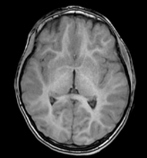
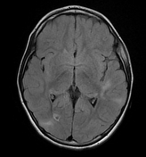
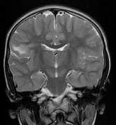
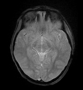
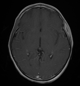
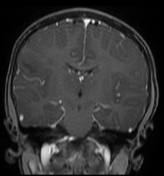
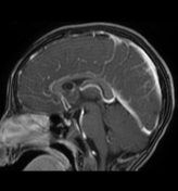
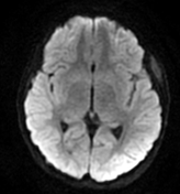
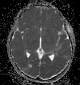
The MRI sequences demonstrate:
- numerous bilateral cortical and subcortical areas of low signal on T1, high signal on T2 and FLAIR with no enhancement on postcontrast sequences in keeping with cortical/subcortical tubers.
- linear bands of high signal T2 and FLAIR are also noted mainly in the left frontal lobe in keeping with radial bands sign.
- multiple small subependymal nodules (hamartomas) along the wall of the lateral ventricles, iso-to high signal to the grey matter on T1, isosignal on T2 and FLAIR with moderate enhancement on postcontrast sequences. Some of them show a low signal on GE (calcified nodules). The largest nodule (8 mm) is located near the right foramen of Monro and appears cystic of low signal on T1, high signal on T2 with peripheral enhancement on postcontrast sequences.
Case Discussion
MRI features most consistent with tuberous sclerosis.


 Unable to process the form. Check for errors and try again.