
Presentation
Background of tuberous sclerosis and intracardiac rhabdomyosarcoma, 2 day history of generalised tonic-clonic seizures.
Patient Data


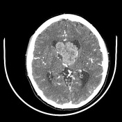
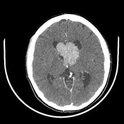

At the level of the third ventricle, extending to the lateral ventricles, a heterogeneous density lesion with well defined, lobulated borders is observed demonstrating heterogeneous enhancement, with resultant dilation of the supratentorial ventricular system.
Diffuse subcortical multiple hypodense, poorly defined foci are also identified and do not demonstrate any enhancement.
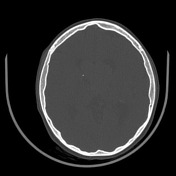
Multiple periventricular subependymal nodules, some with pinpoint calcifications.
Bilateral Galassi I arachnoid cysts and left frontal arachnoid cyst.
Mega cisterna magna.
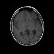
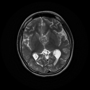
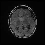
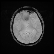
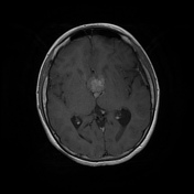
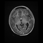
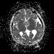
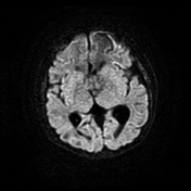
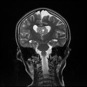
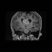
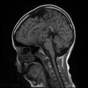
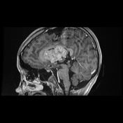

At the level of the third ventricle extending to the lateral ventricles, a lesion with defined lobulated edges is observed, isointense in T1 and T2, hyperintense in FLAIR with heterogeneous post-contrast enhancement, without diffusion restriction. There is associated supratentorial hydrocephalus.
Periventricular subependymal hamartomas with blooming artifact on gradient echo.
Multiple poorly defined areas are identified at the subcortical level, hyperintense on T2 and FLAIR, of diffuse distribution that does not enhance on post contrast sequences.
Bilateral Galassi I arachnoid cysts and left frontal arachnoid cyst.
Mega cisterna magna.
Case Discussion
A 10-year-old patient with a history of tuberous sclerosis and intracardiac rhabdomyosarcoma, consultation for a 2-day clinical picture consisting of generalised tonic-clonic seizures.
Taking into account the clinical history and the CT and MRI findings, it is concluded that it is a typical case of tuberous sclerosis and subependymal giant cell astrocytoma.


 Unable to process the form. Check for errors and try again.