Presentation
A young female came with a history of blurred vision, recurrent muscle weakness episodes on a right hand and foot and focal seizures since 2 weeks. No past medical history.
Patient Data
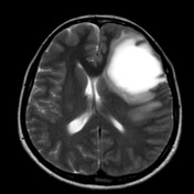

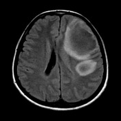

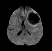

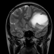

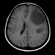

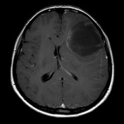

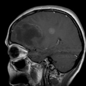

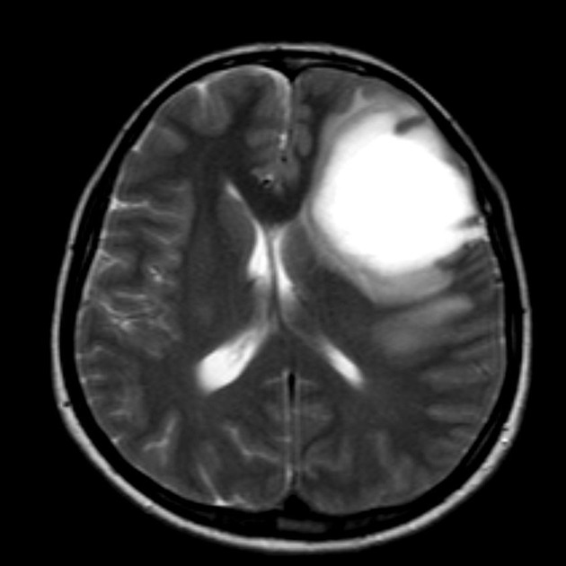
MRI demonstrates two “tumour-like” oval lesions in the left frontoparietal region with extension to centrum semiovale. The lesions are peripherally hypointense and centrally hyperintense on T2WI, show suppression with mild hypointensity on FLAIR and hypointense on T1WI. The margins of lesions show peripherally restricted diffusion on DW images and after contrast administration shows incomplete or open ring enhancement. There is perilesional oedema with mass effect and mild shift midline structures.
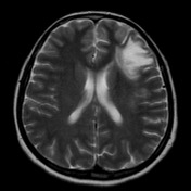

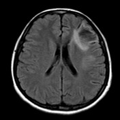

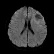

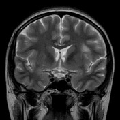

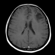

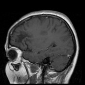

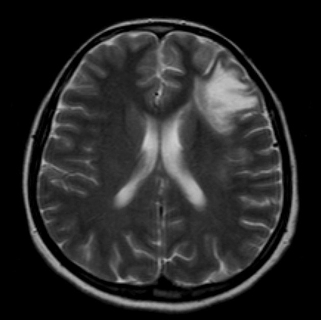
The post-treatment with high dose IV corticosteroids MRI performed after a month.
Post-treatment MRI shows decreasing lesions size, without enhancement of lesions and decrease in the volume of perilesional oedema.
There was marked clinical improvement after high dose IV corticosteroids.
Findings suggestive with tumefactive demyelination.
Case Discussion
Tumefactive demyelinating lesions are an aggressive form of demyelination. There are focal zones of demyelination in the central nervous system and they often mimic a neoplasm.
Most patients with tumefactive demyelinating lesions do respond to steroid therapy.


 Unable to process the form. Check for errors and try again.