
Presentation
The patient was presented with sudden onset of dizziness and double vision three days prior to admission.
Patient Data

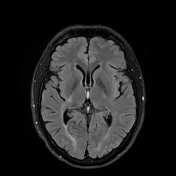
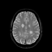
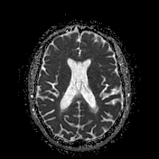
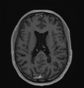
There is a ring-shaped area of high signal on DWI periventricularly, corresponding to the body of the left lateral ventricle near the posterior horn, without corresponding low signal on ADC. A similar but smaller change is also identified next to the anterior horn of the right lateral ventricle. These changes display high signal on the T2 FLAIR sequence, with a surrounding rim or capsule/oedema, particularly around the lesion on the left side. The ventricular system appears midline and age-appropriate. A ring enhancement is seen on T1 with contrast.
There is also a right sided non-enhancing lesion on T2, which is interpreted as a chronic plaque, which also supports the diagnosis (through dissemination in time).
Case Discussion
Tumefactive multiple sclerosis (MS), also known as Balo’s concentric sclerosis or Balo’s disease, is a rare form of MS characterised by mass-like, often large lesions in the brain that can mimic brain tumours on imaging 1,2. These lesions can cause neurological symptoms such as headaches, double vision, balance problems, seizures, motor deficits, or cognitive changes.
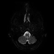
This patient had a previous history of amphetamine and cannabis use and active alcohol overuse before admission. On examination, the patient had interocular ophthalmoplegia. An MRI (TCI protocol) was done to exclude ischaemic changes but revealed ring-shaped changes around the lateral ventricle.
The patient has a family history of multiple sclerosis, as both his mother and grandmother are affected by the condition. The patient had also experienced an episode of slurred vision that lasted for two days, with spontaneous full remission.
The patient initially presented with binocular double vision, both vertical and horizontal, accompanied by dizziness. Upon further questioning, the patient reported experiencing a burning sensation across the right side of the face almost every morning, which was initially attributed to stress, but it was later considered a part of the diagnosis of multiple sclerosis 3.
Following a thorough diagnostic workup, including a lumbar puncture, a spinal cord MRI, blood tests and follow-up brain MRI that showed a ring-shaped area of high signal on DWI periventricularly 4. The patient was diagnosed with multiple sclerosis and started on Ocrelizumab therapy.


 Unable to process the form. Check for errors and try again.