
Presentation
Bleeding.
Patient Data
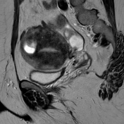

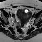
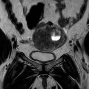
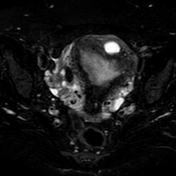
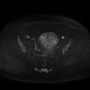
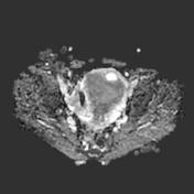
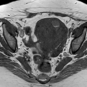
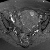
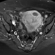
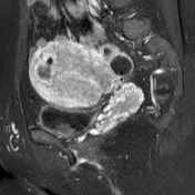

Diffusely enlarged uterus with indistinct junctional zone with multiple myometrial small haemorrhagic foci. It shows a well defined cystic lesion within the fundus which shows a dependent high signal layer and a nondependent intermediate signal layer surrounded by isointense myometrium.
Case Discussion
The case shows both the common diffuse form of adenomyosis and the rare form of cystic adenomyosis.
Cystic adenomyosis is a rare variant of adenomyosis and is believed to be the result of repeated focal haemorrhages resulting in cystic spaces filled with altered blood products.
On MRI it appears as complex cystic lesion showing the following:
It elicits high signal on T1WI and intermediate to high signal on T2WI secondary to haemorrhagic or proteinaceous content
a fluid-fluid level may be seen
the inner cyst wall may present with a thin rim of haemosiderin deposition eliciting persistent low signal on both T1WI and T2WI
lesions are surrounded by T2-hypointense myometrium due to reactive hypertrophy
DDx includes fibroid with haemorrhagic degeneration.
Case courtesy of Dr. Fath Allah Awad. MD of Radiodiagnosis.


 Unable to process the form. Check for errors and try again.