
Presentation
Incidental finding on chest CT.
Patient Data
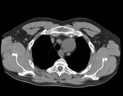

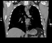

Noncontrast CT of the chest shows two oval, somewhat fusiform masses located in the superior/middle mediastinum that exert mass effect and do not invade adjacent vessels and aerodigestive structures.
Both lesions are similar in appearance, exhibiting relatively circumscribed margins and internal low density suggesting possible composition of soft tissue and fatty elements. There is the suggestion of continuation with a thin linear soft tissue structure superior/inferior to each mass, best appreciated on coronal view.
The superior mass is located anterior to the proximal left subclavian artery at the level of the thoracic inlet. The inferior mass is located along the left tracheoesophageal groove.
Case Discussion
Imaging features of oval/fusiform circumscribed lesion with noninvasive growth pattern and low internal density are highly suggestive of peripheral nerve sheath tumour. In this case, the lesions appear to be in continuation with a nerve, essentially clinching the diagnosis. The location of each lesion suggests the nerves of origin are vagus (superior mass) and recurrent laryngeal nerve (inferior mass).
The lesion underwent biopsy and histologic evaluation confirmed schwannoma. Given the multiplicity of findings, recommendation was made for genetic testing for neurofibromatosis/schwannomatosis.


 Unable to process the form. Check for errors and try again.