
Presentation
Headaches with nausea/vomiting and dizziness.
Patient Data
Age: 55 years
Gender: Male
From the case:
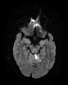
Vermian infarct


Show annotations
Download
Info

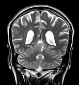
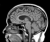
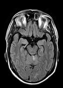
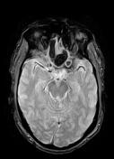
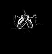
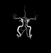
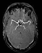
Areas of low signal on T1, high T2 and FLAIR with restricted diffusion involving mainly the superior vermis (superior cerebellar artery territory) more on the left. No hemorrhagic component was seen on the GE sequence. No evidence of arterial occlusion on the MRA 3D-TOF.
Moderate cerebral volume loss.
Case Discussion
MRI features of acute vermian infarct in the superior cerebellar artery territory.


 Unable to process the form. Check for errors and try again.