
Presentation
Patient presented with right-sided weakness, slurred speech, and a decreased level of consciousness.
Patient Data

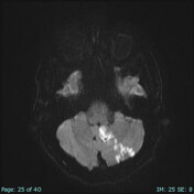
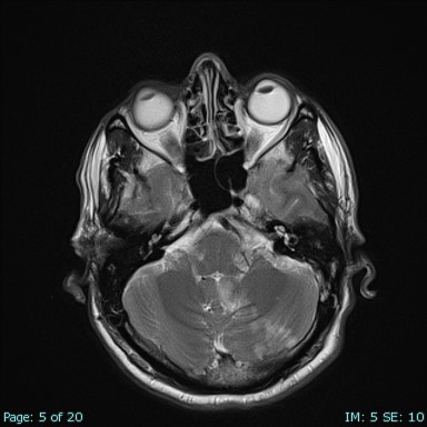
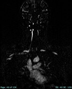
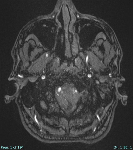
There is an area of restricted diffusion involving the inferomedial side of the left cerebral hemisphere, including the vermis, measuring about 5 x 1 cm and representing acute infarction.
There was no haemorrhagic transformation.
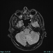
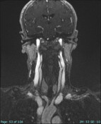
Loss of signal void in the left vertebral artery suggests thrombosis.
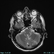
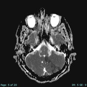
Multiple bilateral T2/FLAIR hyperintensities were seen involving the periventricular and subcortical white matter without diffusion restriction or mass effect,
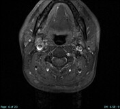
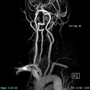

The left vertebral artery from its origin up to the C3/C4 level is not opacified, showing circumferential high T1/T2 fat sat signal changes with a crescent appearance at the C3/C4 level.
Distally, it appears opacified but narrowed compared to the right side.
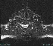
The common carotid, internal, and external carotid arteries bilaterally appear normal without significant stenotic or obstructive pathology.
The right vertebral artery appears normal.
The terminal portions of both internal carotid arteries and basilar arteries and their main branches appear normal.
Angiography was done, and this MRA was after it and shows:
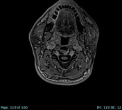
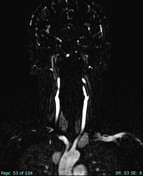
The proximal part of the left vertebral artery is faintly visible; however, there is no opacification of a long segment of the left vertebral artery about 5.3 cm in CC dimension extending from C6/C3 level.
However, the rest of the left vertebral artery appears well opacified, without an obvious luminal defect or cleft.
The right vertebral artery appears normal.
The common carotid, internal, and external carotid arteries bilaterally appear normal without significant stenotic or obstructive pathology.
Case Discussion
This patient came with right-sided weakness and slurred speech; left cerebral hemisphere acute infarction with no visualisation of the left vertebral artery; MRA and carotid MRA were done and showed no opacification of the left vertebral artery from its origin up to the C3/C4 level; however, distally, it appears opacified but narrowed compared to the right side.
After angiography, only the proximal part of the left vertebral artery is faintly opacified.


 Unable to process the form. Check for errors and try again.