
Presentation
Progressive headache.
Patient Data
Age: 45 years
Gender: Female
From the case:
Vestibular schwannoma

Download
Info

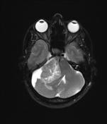
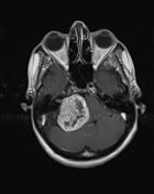
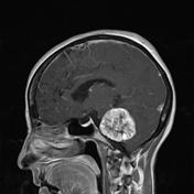
There is a large heterogenous extra axial mass in the right CP angle which is hyperintensity in T2WI and isointensity to gray matter in T1WI. The mass demonstrates heterogenous avid and displaces and compressed the pons, midbrain and fourth ventricle.
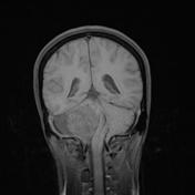
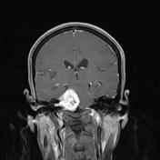
Continuation of the tumor with the vestibular nerve is apparent in axial and coronal post contrast images.
Incidental finding of a partial empty sella.
Case Discussion
The nature of the lesion was pathologically confirmed to be an acoustic schwannoma.


 Unable to process the form. Check for errors and try again.