
Presentation
Headache and fever. CSF lymphocytosis on lumbar puncture.
Patient Data

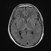
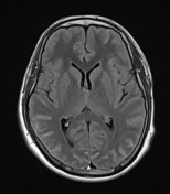
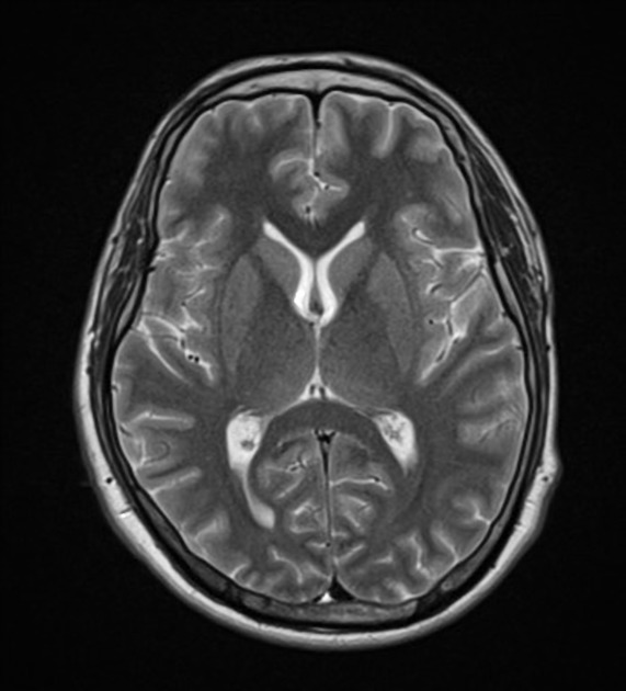
Normal CT study. No intracranial haemorrhage or acute infarction. Normal grey white matter differentiation. No abnormal contrast enhancing lesions. The basal cisterns are normal. Ventricular and sulcal size are age appropriate. No extra-axial abnormality.


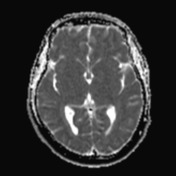
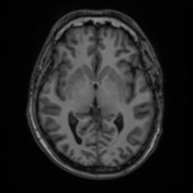
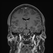
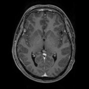
Although not visible on post-contrast T1 and pre-contrast FLAIR, there is extensive smooth thin sulcal enhancement on the more sensitive post-contrast FLAIR sequence.
Case Discussion
Although not visible on post-contrast T1 and pre-contrast FLAIR, there is extensive smooth thin sulcal enhancement on the more sensitive post-contrast FLAIR sequence. The CT is normal and the remainder of the MRI is normal.
Diffuse leptomeningeal enhancement is in-keeping with meningitis. The lumbar puncture demonstrated clear and colourless CSF with a CSF lymphocytosis. The gram stain and culture were negative. The differential in this case includes viral meningitis, or less likely TB or fungal meningitis.


 Unable to process the form. Check for errors and try again.