
Presentation
Non-tender abdominal lump.
Patient Data


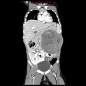

A very large heterogeneously enhancing soft-tissue density mass appears to arise from the left kidney and results in hydronephrosis of the collecting system which is located posterior to the mass. The mass is well circumscribed without evidence of direct extension in adjacent organs/structures. No convincing nodal enlargement.
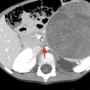
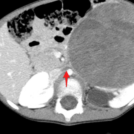
Low-density filling defect (red arrow) in the left renal vein suggesting extension of the tumour.
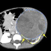
Claw sign is strong evidence that the mass arises from the kidney. Note how the renal parenchyma is stretched around the mass at the margins (yellow arrows).
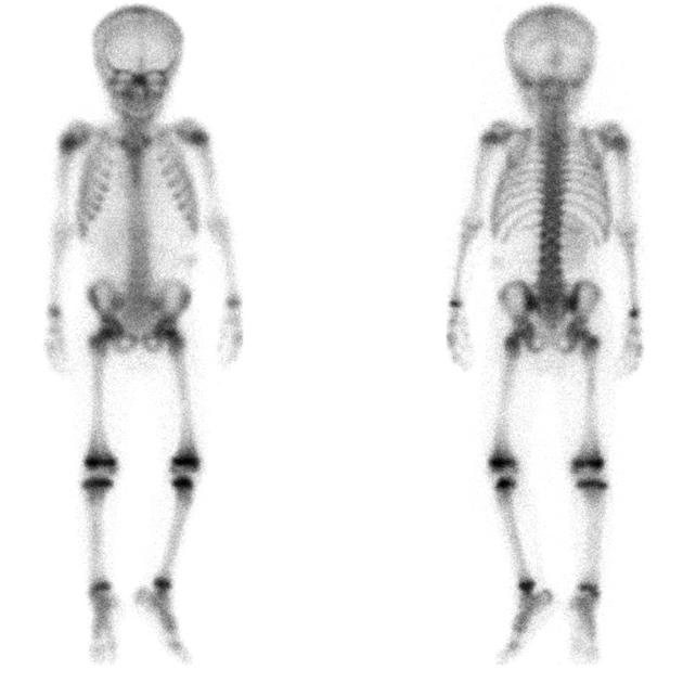
Bone scan (bone phase) using 99mTc-methylene diphosphonate (MDP) demonstrates no abnormal uptake in the skeleton.
Case Discussion
This patient went on to have a biopsy which confirmed the diagnosis of Wilms tumour.


 Unable to process the form. Check for errors and try again.