MRI
Axial images through the skull base show a cystic lesion just lateral to the left inferior cerebellar peduncle, this cystic lesion has CSF density and no evidence of diffusion restriction. It's suggestive of an arachnoid cyst.
This cystic lesion is exerting mass effect on the left facial and vestibulocochlear nerves before entering the internal auditory canal and on the left glossopharyngeal and vagus nerves before entering the jugular foramen.
The scalp shows a midline lesion in association with suggestive bony defect, through which multiple vessles are seen. This appearance and location suggest a sinus pericranii, further evaluation by contrast-enhanced study is recommended.
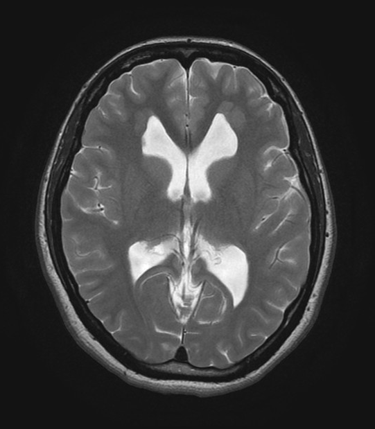
Extensive periventricular gray matter nodular heterotopia is seen with indentation in lateral ventricles walls, extensive bilateral heterotopic gray matter are also seen in the frontal subcortical region extending into the frontal horns of the lateral ventricles.


