Acquired cholesteatoma
- Integral Diagnostics, Shareholder (ongoing)
- Micro-X Ltd, Shareholder (ongoing)
Updates to Article Attributes
Acquired cholesteatomas are far more common than congenital cholesteatomas and are almost always closely related to the tympanic membrane and and pneumatised portion of the temporal bone from which most are thought to arise 9.
Cholesteatomas occur far more commonly in the middle ear than in the external auditory canal. This article relates to middle-ear acquired cholesteatomas. See the external auditory canal cholesteatoma article for details on that entity.
Epidemiology
Acquired cholesteatomas make up 98% of all middle ear cholesteatomas 9.
Clinical presentation
The vast majority of acquired cholesteatomas develop as a result of due to chronic middle ear infectionsotitis media and are usually associated with perforation of the tympanic membrane. Clinical presentation usually consists of conductive hearing loss, often with purulent discharge from the ear 6.
Patients may also present due to one of many complications, which include:
cochlear fistula:
lessless commonfacial nerve dysfunction, including the rare inflammatory neuroma of the facial nerve
extension through the inner ear into the internal acoustic meatus
leadingleading to deafnessextension into the middle cranial fossa
withwith possible meningitis,cerebral abscess, etc.extension into the petrous apex(rare) with similar complications to petrous apicitis
Pathology
Cholesteatomas are composed of densely packed desquamated keratinising squamous cells, arising from a peripheral shell of inward-facing epithelium. As cells mature, they continue to be shed into the mass, resulting in slow growth 1-3.
Aetiology
There are four hypotheses that relate to the formation of cholesteatomas; all all may be true 1,6:
-
invagination/negative pressure
probably the most common cause
results from Eustachian tube dysfunction and tympanic membrane retraction, with debris and keratin eventually obstructing the neck of the retraction
-
invasion/migration
in the setting of a previous perforation
keratinised cells 'invade' the middle ear through the perforation
-
basal cell hyperplasia and papillary ingrowth
: invasive hyperplasia of the basal cell layer of the tympanic membrane as a result of infection
-
metaplasia
: as a result of chronic irritation from middle ear infection
Location
There are a number of subtypes of acquired cholesteatoma, classified by location 11:
-
middle ear cholesteatoma cholesteatoma (most common)
pars flaccida type
pars tensa type
Radiographic features
CT
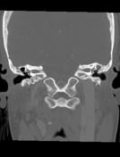
CT is the modality of choice for diagnostic assessment of cholesteatomas, due to its ability to demonstrate the bony anatomy of the temporal bone in exquisite detail.
CholesteatomasCholesteatomas appear as regions of soft-tissue attenuationtissue density, exerting mass effect and resulting in bony erosion, with the latter the hallmark of cholesteatoma 11.Findings depend on the part of the tympanic membrane the middle ear cholesteatoma arises from:
-
pars flaccida
(82%type (more common)superior extension: most common, it expands into Prussak space, eventually eroding the scutum
and, displacing the ossiclesmediallymedially, and eroding the aditus ad antrum posteriorly (sometimes forming a common cavity involving the epitypmanum and aditus ad antrum) 11inferior extension: less common, but more frequently seen in childrenref
-
pars tensa type
MRI
Although MRI is unable to adequately delineate bony anatomy, it can potentially distinguish non-specific opacification from cholesteatomas. It is particularly useful in the postoperative setting when CT may be indeterminate, since granulation tissue, scarring and recurrent cholesteatoma may all appear similar 2.
Cholesteatomas demonstrate:Signal characteristicss
T1:
lowlow signalT2:
highhigh signalT1 C+ (Gd):
nono enhancementDWI:
diffusiondiffusion restriction
Diffusion-weighted imaging is particularly useful when distinguishing a cholesteatoma from other middle ear masses. It is the only entity that demonstrates high signal intensity on DWI. However, the sequence is prone to artefact and care must be taken how the sequence is performed and interpreted 2. Non-echo planar DWI is superior for the diagnosis of cholesteatoma and is therefore preferred if it is available 8. DWI (especially non-EPI DWI) is particularly useful in cases of suspected post-surgical recurrence.
The mechanism responsible for a high signal on DWI remains somewhat uncertain but is thought to represent either T2 shine-through
alonealone or in combination with true restricted diffusion 2-4.Treatment and prognosis
Surgical excision is curative. However, recurrence is not uncommon because the lesion is often difficult to remove completely.
Differential diagnosis
The differential is of a middle ear mass
includeswith bony erosion 11:-
high T1 signalno enhancementno restriction diffusion
mucoid impactionother tumours, e.g. rhabdomyosarcoma, squamous cell carcinoma, metastasis, giant cell tumour
chronic otitis media: characterised by tympanic membrane retruction
Cholesteatoma is difficult to differentiate on CT from other middle ear masses without bony erosion with MRI helpful in differentiatin cholesteatoma from other entities 11:
cholesterol granuloma: high T1 signal, no enhancement, no restriction diffusion
granulation tissue/fibrosis/scar tissue
post-inflammatory ossicular fixation
mucosal oedema
Post-operative
Following resection of a cholesteatoma, the differential for a soft-tissue middle ear mass includes the entities above, but is usually restricted to three entities 2,10:
-
recurrent cholesteatoma
low T1 signal
no enhancement
increased signal on DWI
-
granulation tissue
intermediate T1 signal
enhancement
low signal on DWI
-
scarring
low T1 and T2 signal
low signal on DWI
Practical points
careful review of areas not visible on otoscopy should be performed including the sinus tympani, facial recess, and anterior epitympanic recess11
-<p><strong>Acquired cholesteatomas</strong> make up 98% of all <a href="/articles/middle-ear">middle ear</a> cholesteatomas and are almost always closely related to the <a href="/articles/tympanic-membrane">tympanic membrane</a> and pneumatised portion of the <a href="/articles/temporal-bone-1">temporal bone</a> from which most are thought to arise <sup>9</sup>. </p><h4>Clinical presentation</h4><p>The vast majority of acquired cholesteatomas develop as a result of chronic middle ear infections and are usually associated with perforation of the tympanic membrane. Clinical presentation usually consists of <a href="/articles/conductive-hearing-loss">conductive hearing loss</a>, often with purulent discharge from the ear <sup>6</sup>. </p><p>Patients may also present due to one of many complications, which include: </p><ul>-<li><p><a href="/articles/labyrinthine-fistula">labyrinthine fistula</a> (<a href="/articles/perilymphatic-fistula">perilymphatic fistula</a>)</p></li>-<li><p><a href="/articles/cochlear-fistula">cochlear fistula</a>: less common</p></li>- +<p><strong>Acquired cholesteatomas</strong> are far more common than <a href="/articles/congenital-cholesteatoma" title="Congenital cholesteatomas">congenital cholesteatomas</a> and are almost always closely related to the <a href="/articles/tympanic-membrane">tympanic membrane</a> and pneumatised portion of the <a href="/articles/temporal-bone-1">temporal bone</a> from which most are thought to arise <sup>9</sup>. </p><p>Cholesteatomas occur far more commonly in the <a href="/articles/middle-ear" title="Middle ear">middle ear</a> than in the <a href="/articles/external-auditory-canal" title="External auditory canal">external auditory canal</a>. This article relates to middle-ear acquired cholesteatomas. See the <a href="/articles/external-auditory-canal-cholesteatoma" title="External auditory canal cholesteatoma">external auditory canal cholesteatoma</a> article for details on that entity. </p><h4>Epidemiology</h4><p>Acquired cholesteatomas make up 98% of all <a href="/articles/middle-ear">middle ear</a> cholesteatomas <sup>9</sup>.</p><h4>Clinical presentation</h4><p>The vast majority of acquired cholesteatomas develop due to <a href="/articles/chronic-otitis-media" title="Chronic otitis media">chronic otitis media</a> and are usually associated with perforation of the tympanic membrane. Clinical presentation usually consists of <a href="/articles/conductive-hearing-loss">conductive hearing loss</a>, often with purulent discharge from the ear <sup>6</sup>. </p><p>Patients may also present due to one of many complications, which include: </p><ul>
- +<li><p><a href="/articles/labyrinthine-fistula">labyrinthine fistula</a> (<a href="/articles/perilymphatic-fistula">perilymphatic fistula</a>)</p></li>
- +<li><p><a href="/articles/cochlear-fistula">cochlear fistula</a>: less common</p></li>
-<li><p>facial nerve dysfunction including the rare <a href="/articles/inflammatory-neuroma-of-the-facial-nerve">inflammatory neuroma of the facial nerve</a></p></li>-<li><p>extension through the inner ear into the <a href="/articles/internal-acoustic-canal">internal acoustic meatus</a> leading to deafness</p></li>-<li><p>extension into the <a href="/articles/middle-cranial-fossa">middle cranial fossa</a> with possible <a href="/articles/leptomeningitis">meningitis</a>, <a href="/articles/cerebral-abscess-1">cerebral abscess</a>, etc.</p></li>-<li><p>extension into the <a href="/articles/petrous-apex">petrous apex</a> (rare) with similar complications to <a href="/articles/petrous-apicitis">petrous apicitis</a></p></li>-</ul><h4>Pathology</h4><p>Cholesteatomas are composed of densely packed desquamated keratinising squamous cells, arising from a peripheral shell of inward-facing epithelium. As cells mature, they continue to be shed into the mass, resulting in slow growth <sup>1-3</sup>. </p><p>There are four hypotheses that relate to the formation of cholesteatomas; all may be true <sup>1,6</sup>:</p><ul>- +<li><p>facial nerve dysfunction, including the rare <a href="/articles/inflammatory-neuroma-of-the-facial-nerve">inflammatory neuroma of the facial nerve</a></p></li>
- +<li><p>extension through the inner ear into the <a href="/articles/internal-acoustic-canal">internal acoustic meatus</a> leading to deafness</p></li>
- +<li><p>extension into the <a href="/articles/middle-cranial-fossa">middle cranial fossa</a> with possible <a href="/articles/leptomeningitis">meningitis</a>, <a href="/articles/cerebral-abscess-1">cerebral abscess</a>, etc.</p></li>
- +<li><p>extension into the <a href="/articles/petrous-apex">petrous apex</a> (rare) with similar complications to <a href="/articles/petrous-apicitis">petrous apicitis</a></p></li>
- +</ul><h4>Pathology</h4><p>Cholesteatomas are composed of densely packed desquamated keratinising squamous cells, arising from a peripheral shell of inward-facing epithelium. As cells mature, they continue to be shed into the mass, resulting in slow growth <sup>1-3</sup>. </p><h5>Aetiology</h5><p>There are four hypotheses that relate to the formation of cholesteatomas; all may be true <sup>1,6</sup>:</p><ul>
- +<li><p>basal cell hyperplasia and papillary ingrowth: invasive hyperplasia of the basal cell layer of the tympanic membrane as a result of infection</p></li>
- +<li><p>metaplasia: as a result of chronic irritation from middle ear infection</p></li>
- +</ul><h5>Location</h5><p>There are a number of subtypes of acquired cholesteatoma, classified by location <sup>11</sup>:</p><ul>
-<p>basal cell hyperplasia and papillary ingrowth</p>-<ul><li><p>invasive hyperplasia of the basal cell layer of the tympanic membrane as a result of infection</p></li></ul>-</li>-<li>-<p>metaplasia</p>-<ul><li><p>as a result of chronic irritation from middle ear infection</p></li></ul>-</li>-</ul><h4>Radiographic features</h4><h5>CT</h5><p>CT is the modality of choice for diagnostic assessment of cholesteatomas, due to its ability to demonstrate the bony anatomy of the temporal bone in exquisite detail. Cholesteatomas appear as regions of soft-tissue attenuation, exerting mass effect and resulting in bony erosion. </p><p>Findings depend on the part of the <a href="/articles/tympanic-membrane">tympanic membrane</a> the cholesteatoma arises from: </p><ul>-<li>-<p><a href="/articles/pars-flaccida">pars flaccida</a> (82%) </p>- +<p>middle ear cholesteatoma cholesteatoma (most common)</p>
-<li><p>superior extension: most common, it expands into <a href="/articles/prussak-space">Prussak space</a>, eventually eroding the <a href="/articles/scutum">scutum</a> and displacing the <a href="/articles/middle-ear-ossicles">ossicles</a> medially</p></li>-<li><p>inferior extension: less common, but more frequently seen in children</p></li>- +<li><p>pars flaccida type</p></li>
- +<li><p>pars tensa type</p></li>
- +<li><p><a href="/articles/external-auditory-canal-cholesteatoma" title="External auditory canal cholesteatoma">external auditory canal cholesteatoma</a> (rare)</p></li>
- +</ul><h4>Radiographic features</h4><h5>CT</h5><p>CT is the modality of choice for diagnostic assessment of cholesteatomas, due to its ability to demonstrate the bony anatomy of the temporal bone in exquisite detail. Cholesteatomas appear as regions of soft tissue density, exerting mass effect and resulting in <a href="/articles/bone-erosion" title="Bony erosion">bony erosion</a>, with the latter the hallmark of cholesteatoma <sup>11</sup>. </p><p>Findings depend on the part of the <a href="/articles/tympanic-membrane">tympanic membrane</a> the middle ear cholesteatoma arises from: </p><ul>
-<p><a href="/articles/pars-tensa">pars tensa</a></p>- +<p><a href="/articles/pars-flaccida">pars flaccida</a> type (more common)</p>
-<li><p>posterosuperior (78%): extends medial to the <a href="/articles/incus">incus</a> and displaces the <a href="/articles/middle-ear-ossicles">ossicles</a> laterally</p></li>-<li><p>anterior and inferior (22%)</p></li>- +<li><p>superior extension: most common, it expands into <a href="/articles/prussak-space">Prussak space</a>, eventually eroding the <a href="/articles/scutum">scutum</a>, displacing the <a href="/articles/middle-ear-ossicles">ossicles</a> medially, and eroding the aditus ad antrum posteriorly (sometimes forming a common cavity involving the epitypmanum and aditus ad antrum) <sup>11</sup></p></li>
- +<li><p>inferior extension: less common, but more frequently seen in children <sup>ref</sup></p></li>
-</ul><h5>MRI</h5><p>Although MRI is unable to adequately delineate bony anatomy, it can potentially distinguish non-specific opacification from cholesteatomas. It is particularly useful in the postoperative setting when CT may be indeterminate, since granulation tissue, scarring and recurrent cholesteatoma may all appear similar <sup>2</sup>. </p><p>Cholesteatomas demonstrate:</p><ul>-<li><p><strong>T1:</strong> low signal</p></li>-<li><p><strong>T2:</strong> high signal</p></li>-<li><p><strong>T1 C+ (Gd):</strong> no enhancement</p></li>-<li><p><strong>DWI:</strong> diffusion restriction</p></li>-</ul><p>Diffusion-weighted imaging is particularly useful when distinguishing a cholesteatoma from other middle ear masses. It is the only entity that demonstrates high signal intensity on DWI. However, the sequence is prone to artefact and care must be taken how the sequence is performed and interpreted <sup>2</sup>. Non-echo planar DWI is superior for the diagnosis of cholesteatoma and is therefore preferred if it is available <sup>8</sup>. DWI (especially non-EPI DWI) is particularly useful in cases of suspected post-surgical recurrence.</p><p>The mechanism responsible for a high signal on <a href="/articles/dwi">DWI</a> remains somewhat uncertain but is thought to represent either <a href="/articles/t2-shine-through">T2 shine-through</a> alone or in combination with true restricted diffusion <sup>2-4</sup>.</p><h4>Treatment and prognosis</h4><p>Surgical excision is curative. However, recurrence is not uncommon because the lesion is often difficult to remove completely. </p><h4>Differential diagnosis</h4><p>The differential of a <a href="/articles/middle-ear-mass">middle ear mass</a> includes:</p><ul>-<p><a href="/articles/cholesterol-granuloma">cholesterol granuloma</a></p>- +<p><a href="/articles/pars-tensa">pars tensa</a> type</p>
-<li><p>high T1 signal</p></li>-<li><p>no enhancement</p></li>-<li><p>no restriction diffusion</p></li>- +<li><p>posterosuperior extension (more common of this type): extends medial to the <a href="/articles/incus">incus</a> and displaces the <a href="/articles/middle-ear-ossicles">ossicles</a> laterally <sup>11</sup></p></li>
- +<li><p>anterior and inferior extension <sup>ref</sup></p></li>
-<li><p>mucoid impaction</p></li>-<li><p><a href="/articles/tympanic-paraganglioma" title="Tympanic paraganglioma">tympanic paraganglioma</a></p></li>- +</ul><h5>MRI</h5><p>Although MRI is unable to adequately delineate bony anatomy, it can potentially distinguish non-specific opacification from cholesteatomas. It is particularly useful in the postoperative setting when CT may be indeterminate, since granulation tissue, scarring and recurrent cholesteatoma may all appear similar <sup>2</sup>. </p><h6>Signal characteristicss</h6><ul>
- +<li><p><strong>T1:</strong> low signal</p></li>
- +<li><p><strong>T2:</strong> high signal</p></li>
- +<li><p><strong>T1 C+ (Gd):</strong> no enhancement</p></li>
- +<li><p><strong>DWI:</strong> diffusion restriction</p></li>
- +</ul><p>Diffusion-weighted imaging is particularly useful when distinguishing a cholesteatoma from other middle ear masses. It is the only entity that demonstrates high signal intensity on DWI. However, the sequence is prone to artefact and care must be taken how the sequence is performed and interpreted <sup>2</sup>. Non-echo planar DWI is superior for the diagnosis of cholesteatoma and is therefore preferred if it is available <sup>8</sup>. DWI (especially non-EPI DWI) is particularly useful in cases of suspected post-surgical recurrence.</p><p>The mechanism responsible for a high signal on <a href="/articles/dwi">DWI</a> remains somewhat uncertain but is thought to represent either <a href="/articles/t2-shine-through">T2 shine-through</a> alone or in combination with true restricted diffusion <sup>2-4</sup>.</p><h4>Treatment and prognosis</h4><p>Surgical excision is curative. However, recurrence is not uncommon because the lesion is often difficult to remove completely. </p><h4>Differential diagnosis</h4><p>The differential is of a <a href="/articles/middle-ear-mass">middle ear mass</a> with bony erosion <sup>11</sup>:</p><ul>
- +<li><p><a href="/articles/tympanic-paraganglioma" title="Tympanic paraganglioma">tympanic paraganglioma</a></p></li>
- +<li><p>other tumours, e.g. <a href="/articles/rhabdomyosarcoma" title="Rhabdomyosarcoma">rhabdomyosarcoma</a>, <a href="/articles/head-and-neck-squamous-cell-carcinoma-overview" title="Squamous cell carcinoma (head and neck)">squamous cell carcinoma</a>, metastasis, giant cell tumour</p></li>
- +<li><p><a href="/articles/langerhans-cell-histiocytosis" title="Langerhans cell histiocytosis">Langerhans cell histiocytosis</a></p></li>
- +<li><p><a href="/articles/chronic-otitis-media" title="Chronic otitis media">chronic otitis media</a>: characterised by tympanic membrane retruction</p></li>
- +</ul><p>Cholesteatoma is difficult to differentiate on CT from other middle ear masses without bony erosion with MRI helpful in differentiatin cholesteatoma from other entities <sup>11</sup>:</p><ul>
- +<li><p><a href="/articles/cholesterol-granuloma">cholesterol granuloma</a>: high T1 signal, no enhancement, no restriction diffusion</p></li>
- +<li><p>granulation tissue/fibrosis/scar tissue</p></li>
- +<li><p>post-inflammatory ossicular fixation</p></li>
- +<li><p>mucosal oedema</p></li>
-</ul>- +</ul><h4>Practical points</h4><ul><li><p>careful review of areas not visible on otoscopy should be performed including the <a href="/articles/sinus-tympani" title="Sinus tympani">sinus tympani</a>, <a href="/articles/facial-recess" title="Facial recess">facial recess</a>, and <a href="/articles/anterior-epitympanic-recess" title="Anterior epitympanic recess">anterior epitympanic recess</a> <sup>11</sup></p></li></ul>
References changed:
- 11. Gulati M, Gupta S, Prakash A, Garg A, Dixit R. HRCT Imaging of Acquired Cholesteatoma: A Pictorial Review. Insights Imaging. 2019;10(1):92. <a href="https://doi.org/10.1186/s13244-019-0782-y">doi:10.1186/s13244-019-0782-y</a> - <a href="https://www.ncbi.nlm.nih.gov/pubmed/31578644">Pubmed</a>
Image 8 CT (bone window) ( update )


 Unable to process the form. Check for errors and try again.