
Perivascular spaces, also known as Virchow-Robin spaces, are fluid-filled spaces that surround small arterioles, capillaries and venules in the brain. Those that surround perforating vessels are frequently seen on routine MRI imaging.
Despite having been described well over a century ago and seen routinely in the majority of MRI studies, significant uncertainty and controversy continue to exist surrounding their anatomy and function.
Their importance can be broadly divided into:
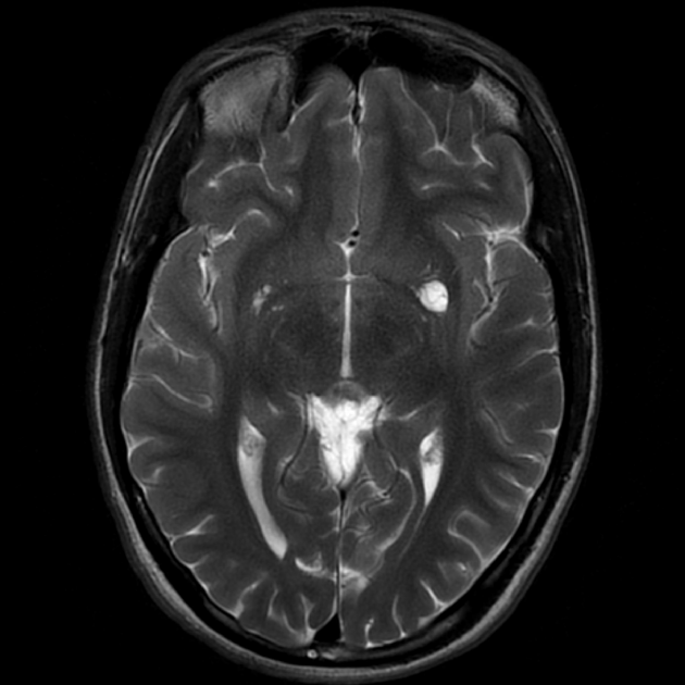
perivascular cysts: larger rounded perivascular spaces mimicking lacunae or cystic lesions
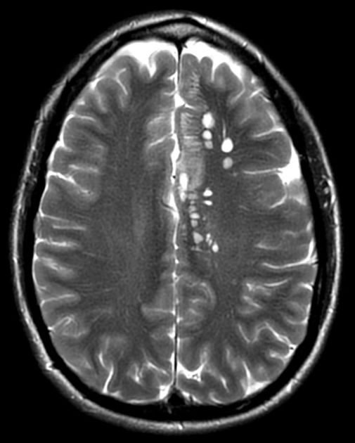
perivascular spaces: smaller linear fluid-intensity structures relevant to neurodegenerative disease (e.g. Alzheimer disease)
On this page:
Terminology
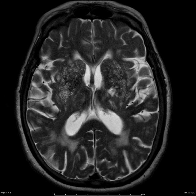
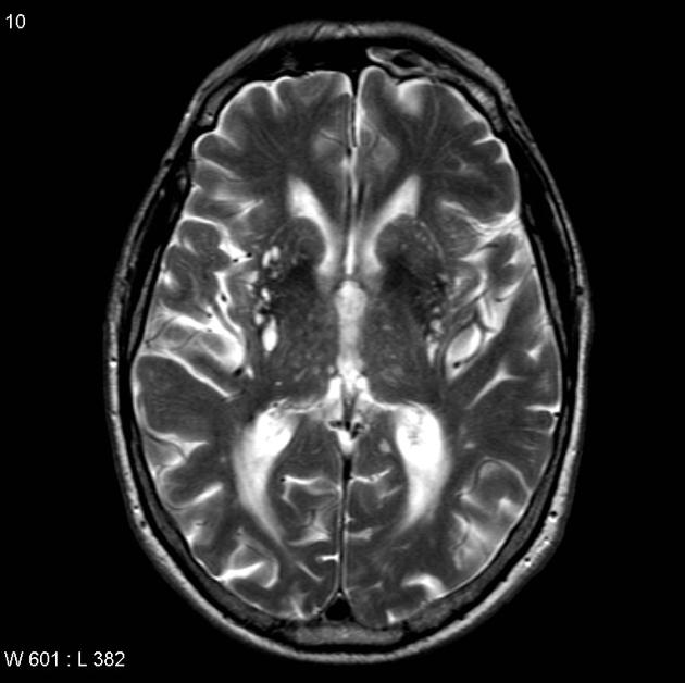
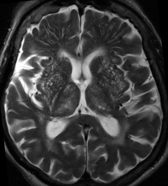
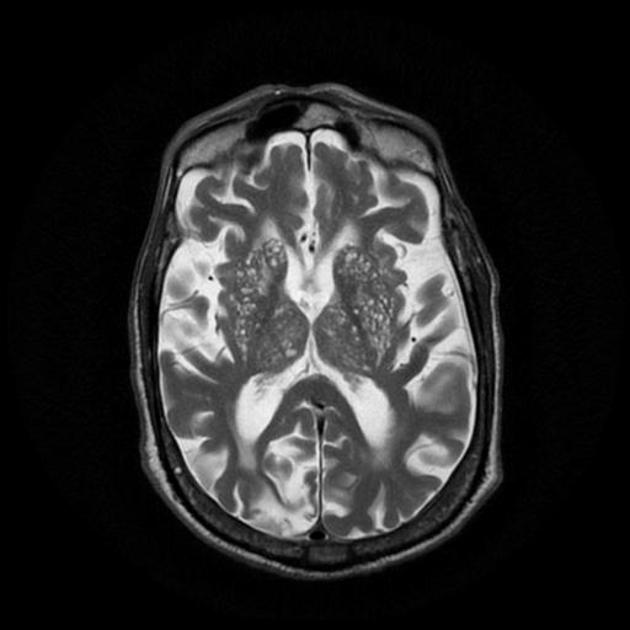
When perivascular spaces are very numerous the brain can have a colander-like appearance, referred to as état criblé (as opposed to numerous lacunar infarcts, sometimes referred to as état lacunaire).
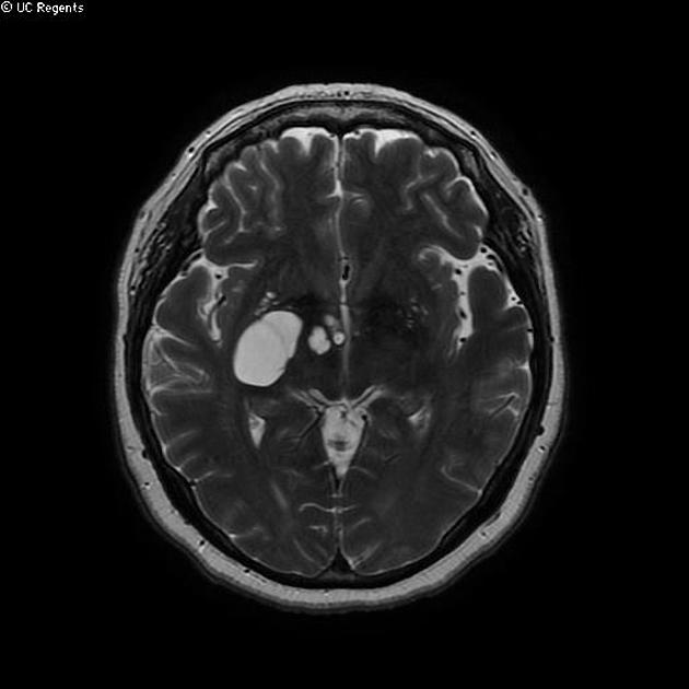
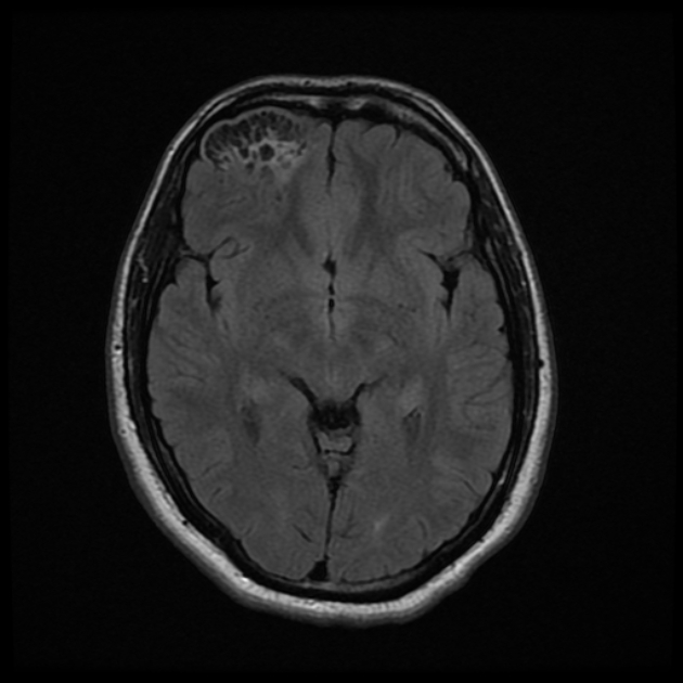
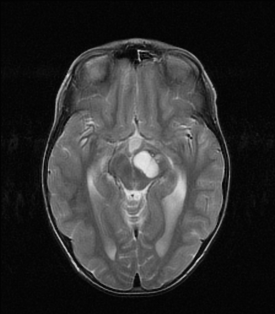
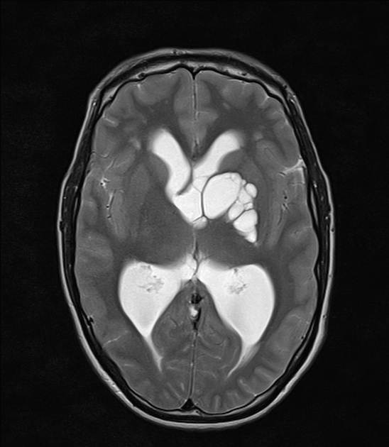
When perivascular spaces are very large, they are referred to as tumefactive perivascular spaces.
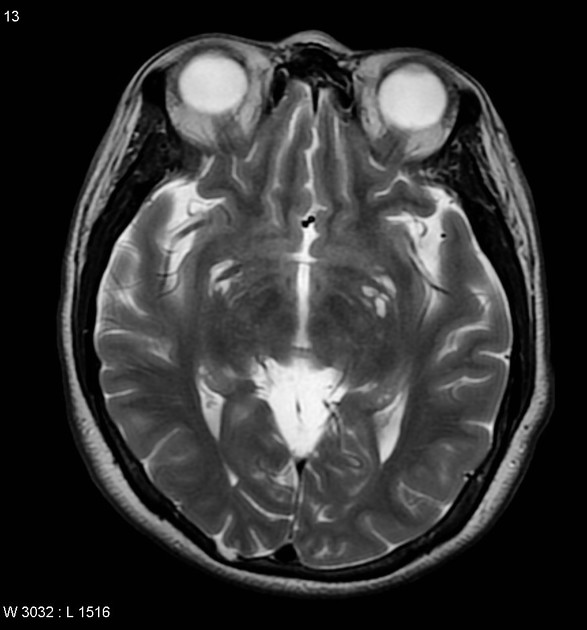
When located in the anterior temporal lobe and related to a vascular loop, they are known as anterior temporal lobe perivascular spaces; however, these likely represent a different entity compared to typical scattered perivascular spaces.
Epidemiology
Perivascular spaces are very common, and increasingly seen with better MRI image resolution. Depending on defining criteria, they are seen in 50-100% of patients 2,3.
Associations
Since they were first described in the 1850s, enlarged perivascular spaces were believed to be entirely incidental findings, mostly significant so as not to be mistaken for a more sinister pathology.
However, in the 2010s, research began to suggest an association between extensive basal ganglia perivascular spaces (état criblé) and changes of chronic microvascular ischemic disease 12-14,20. This is related to the observation that, although a few scattered perivascular spaces are a nearly ubiquitous imaging finding, the number and prominence of these spaces increases with aging, along with other findings of microvascular disease, e.g. periventricular white matter lesions and lacunar infarcts. The association remains controversial 14.
Similarly, the association of enlarged perivascular spaces with subsequent development of dementia has been reported but variably so 20.
Enlarged perivascular spaces have also been reported with greater frequency in a variety of settings, albeit generally in smaller size cohorts 1,4,11,20:
autism spectrum disorder
-
some neurodegenerative conditions, e.g. Parkinson disease, Alzheimer disease
not established markers of disease
some muscular dystrophies
Clinical presentation
Perivascular spaces are normal anatomical structures. Even when enlarged they are almost invariably asymptomatic, even when quite large. Rarely, they can cause mass-effect and can result in obstructive hydrocephalus.
Some postulated that an increased number of perivascular spaces may be a marker of evolving neurodegenerative diseases, including Alzheimer disease and Parkinson disease 18. The relationship between prominent perivascular spaces and disease remains poorly understood 18.
Pathology
Perivascular spaces are normal, usually microscopic structures, that consist of a single or double layer of invaginated pia and basement membrane - depending on location - surrounding small cerebral blood vessels 8,14,20.
Perivascular spaces that surround perforating arteries at the base of the skull, extending into the basal ganglia and internal capsule appear to communicate directly with the subarachnoid space, and thus are presumably filled with CSF, whereas those that surround perforating vessels in the subcortical white matter appear to remain subpial and are thus presumably filled with interstitial fluid 20,21.
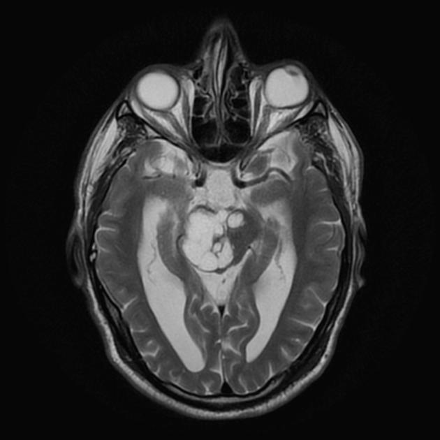
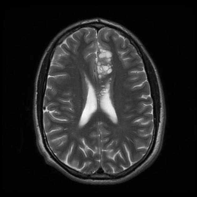
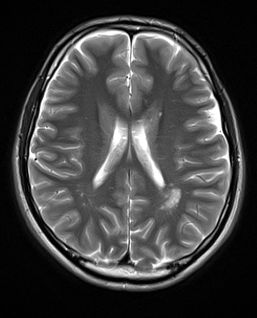
Perivascular spaces that are visible on imaging take on two forms, either thin linear regions, most commonly seen in the centrum semiovale or more oval or cystic appearing spaces, typically <5 mm in diameter, most often seen at the base of the brain. These cystic perivascular spaces can reach much larger sizes, so-called "giant" perivascular space or tumefactive perivascular space, and can exert enough mass effect to be symptomatic 1.
Classification
Perivascular spaces are divided into three main types, although in 2020 a fourth type was described 4,19:
type 1: located in the area supplied by the lenticulostriate arteries entering the basal ganglia
type 2: located in the area supplied by the perforating medullary arteries as they enter the cortical grey matter
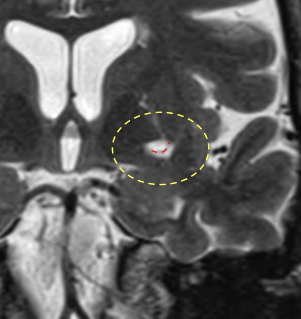
type 3: located in the midbrain
type 4: temporal pole, insular - see anterior temporal lobe perivascular spaces 9,10,19
Radiographic features
Perivascular spaces and cysts are filled with fluid similar to CSF in an appearance on all imaging modalities and sequences. There is no clear cutoff between a visible perivascular space and a cystic perivascular space as they appear to exist along a continuum ref.
Perivascular spaces are seen as linear regions, most frequently seen in the basal ganglia long perforating arteries and in the subcortical white matter ref.
Perivascular cysts are rounded, sometimes seen surrounding a vessel with smooth margins ref.
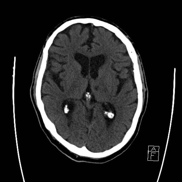
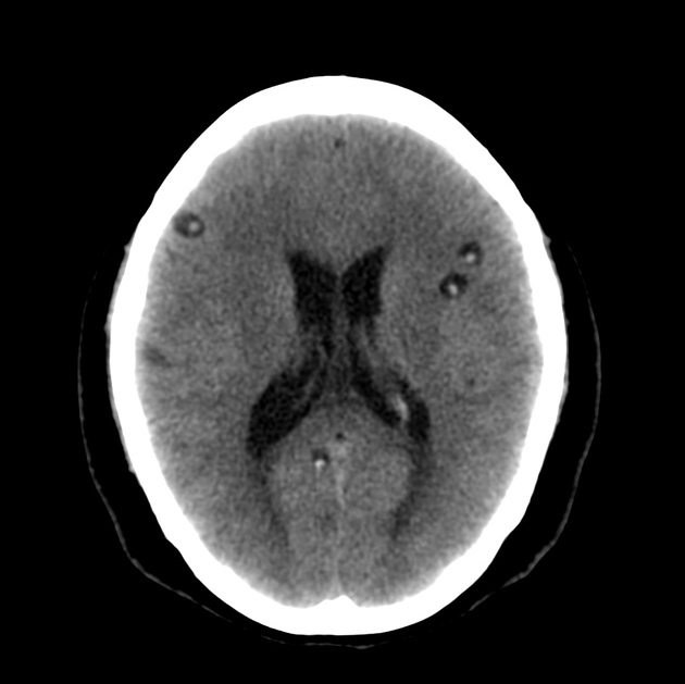
CT
well-circumscribed fluid-density spaces
no enhancement
no calcification
CT angiography occasionally demonstrates a traversing vessel
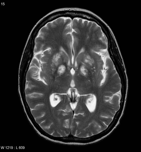
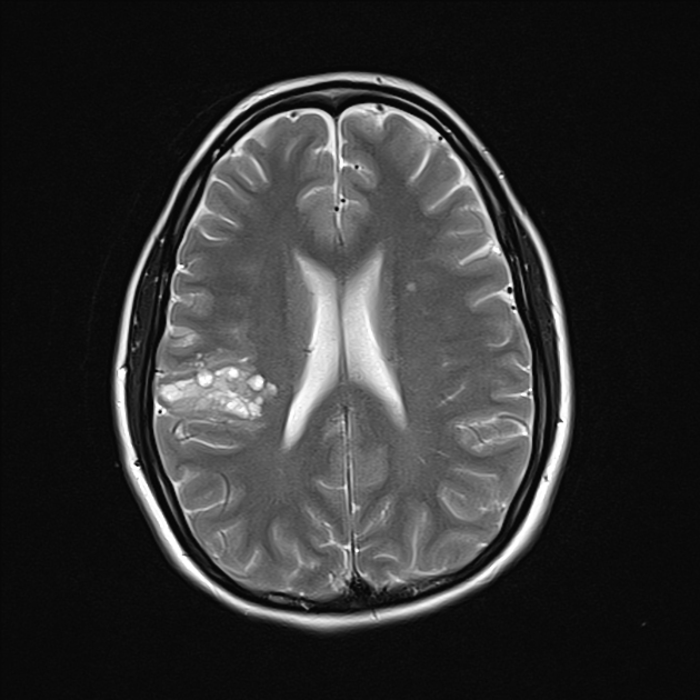
MRI
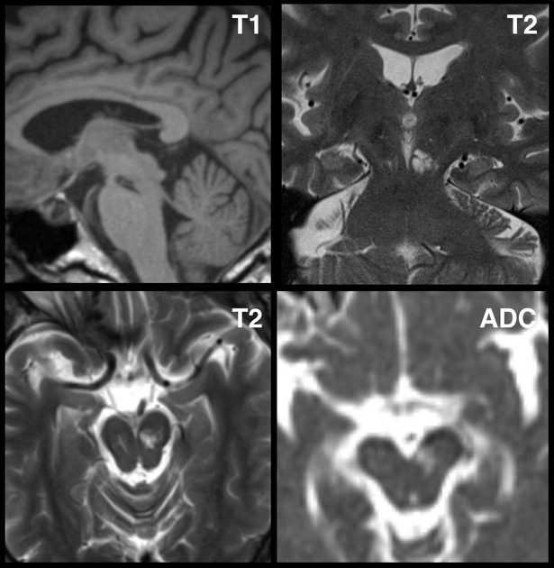
Perivascular spaces follow CSF signal on all pulse sequences 7. When small, the adjacent white matter is normal, thus helping to distinguish perivascular spaces from lacunar infarcts, which have surrounding gliosis (best seen on T2 FLAIR sequence).
In a minority of cases, especially when they are large, a thin increased T2-signal halo may be seen. Usually, they will have a positive mass effect. On T2 sequences, a traversing vessel is sometimes seen.
The exception to the 'no surrounding high T2 signal' rule is anterior temporal lobe perivascular spaces 9,10.
History and etymology
Perivascular spaces were first described by Durand-Fardel (1842) - who described état criblé - and Pestalozzi (1849) 20.
Virchow-Robin spaces are named after German pathologist Rudolf Virchow (1821–1902) 15 and French anatomist Charles-Philippe Robin (1821–1885) who described them further in 1851 and 1859, respectively 16,20. Interestingly, Virchow and Robin disagreed on whether or not these spaces directly communicated with the subarachnoid space. Over a century and a half later, this remains an unresolved question 20.
Differential diagnosis
For small perivascular spaces, consider:
-
lacunar infarcts and striatocapsular infarcts
a rim of gliosis seen best on FLAIR 8
neutral or negative mass effect
typically upper two-thirds of basal ganglia (due to infarcts of perforating end arteries)
-
cyst with a dot sign
enhancement
calcification
CNS cryptococcosis: if multiple
For giant perivascular spaces consider:
-
cystic neoplasms 1,8
they rarely exhibit intensity similar to CSF on all MRI sequences
non-neoplastic neuroepithelial cyst(s)


 Unable to process the form. Check for errors and try again.