
Cases
By sharing our collective experience through interesting and classic patient cases, we can make a real difference in how people are imaged and diagnosed. Each case belongs to a contributing member and all cases are reviewed by our dedicated editors to ensure they reach quality standards and abide by privacy guidelines. Cases can public or unlisted and then be viewed directly or added to articles, playlists or multiple choice questions. Find out more about cases.


58,611 results found
Case
Empyema necessitans

Published
13 May 2024
68% complete
CT
Case
Achilles tendon tear
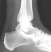
Published
13 May 2024
94% complete
X-ray
Case
Calcific tendinitis of the peroneus longus

Published
13 May 2024
77% complete
MRI
Case
Spinal intradural lipoma

Published
13 May 2024
95% complete
CT
MRI
Case
Unknown pregnancy (CT)

Published
12 May 2024
89% complete
CT
Case
Pelviureteric junction obstruction and MCDK

Published
12 May 2024
81% complete
MRI
CT
Case
Peripelvic cysts and pseudohydronephrosis

Published
12 May 2024
95% complete
Ultrasound
MRI
Case
Craniotomies

Published
12 May 2024
19% complete
Diagram
Case
Paget disease

Published
12 May 2024
69% complete
X-ray
Case
Bilateral giant renal angiomyolipomas

Published
12 May 2024
95% complete
CT
Case
Cavernous sinus meningioma

Published
12 May 2024
77% complete
MRI
Case
Non-small cell lung carcinoma

Published
11 May 2024
90% complete
Pathology
X-ray
Case
ACL and PCL reconstruction

Published
11 May 2024
94% complete
X-ray
Case
ACL reconstruction knee radiograph

Published
11 May 2024
94% complete
X-ray
Case
Autosomal recessive polycystic kidney disease (ARPKD) - antenatal

Published
11 May 2024
75% complete
Ultrasound
Case
Meningioma

Published
11 May 2024
68% complete
CT
Case
Anorectal malformation

Published
11 May 2024
91% complete
X-ray
Case
Disseminated visceral fungal abscesses

Published
10 May 2024
92% complete
CT
Ultrasound
Case
Paraovarian cyst with ovarian torsion

Published
10 May 2024
75% complete
Ultrasound
Photo
Case
Superior mesenteric artery and replaced right hepatic artery thrombosis

Published
10 May 2024
92% complete
CT
ADVERTISEMENT: Supporters see fewer/no ads


 Unable to process the form. Check for errors and try again.