
Adrenal gland trauma most commonly results from blunt force trauma.
On this page:
Epidemiology
Adrenal gland trauma is present on 1-2% of CT imaging in blunt trauma although the occurrence is thought to be much higher as injury has been demonstrated at 28% in one autopsy series 1-4.
The right adrenal gland is more commonly affected than the left with a ratio of 3-4:1 2.
Pathology
Adrenal haemorrhage is the most common injury to the adrenal gland and is thought to be a result of direct compression or increased venous pressures from IVC compression. Laceration of the adrenal gland is less common 2,3.
Associations
Isolated adrenal gland trauma is uncommon (<5% of all adrenal trauma 4) and associated injuries include 1,2:
liver, spleen and/or renal laceration
rib, pelvic or spinal fractures
head injury
Adrenal gland traumatic haemorrhage may also present with 1:
posterior pararenal space haemorrhage
IVC compression
thickening of the diaphragmatic crus
Radiographic features
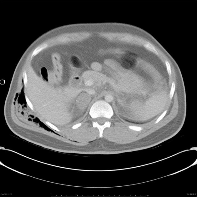
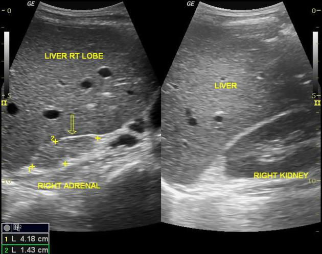
CT
haematoma presents as a well-defined nodular mass, within either the body or the limb with a density of 50-80 HU 1,2
diffuse enlargement or replacement of the adrenal gland with less well-defined borders can also be seen and most often relates to laceration 1,2
periadrenal fat stranding is often present 1
MRI
T1: haematoma is isointense to muscle, liver, renal cortex
T2: haematoma is very hyperintense; hyperintense fat stranding 3
Treatment and prognosis
Adrenal gland trauma is important to recognise as mortality is twice as high in blunt trauma patients with adrenal gland injury than without 2. Management is often conservative but may vary depending on haemodynamic stability and the presence of active bleeding 6.
Complications include 1,4:
acute adrenal insufficiency (if bilateral)
delayed haemorrhage
pseudocyst formation
thrombus from IVC compression (rare)
Differential diagnosis
Differentials to consider include:
pre-existing adrenal mass
haemorrhage into the existing adrenal tumour
adrenal gland hyperenhancement: usually bilateral with preserved adrenal gland morphology and in the setting of hypotension
Practical points
The presence of a discrete adrenal mass in the context of blunt trauma without injury to other abdominal organs or CT features of injury warrants follow-up investigation 1,4.


 Unable to process the form. Check for errors and try again.