Citation, DOI, disclosures and article data
Citation:
St-Amant M, Knipe H, Khalighinejad P, et al. Adrenal washout. Reference article, Radiopaedia.org (Accessed on 29 Mar 2025) https://doi.org/10.53347/rID-34779
Disclosures:
At the time the article was last revised Henry Knipe had the following disclosures:
- Micro-X Ltd, Shareholder (past)
These were assessed during peer review and were determined to
not be relevant to the changes that were made.
View Henry Knipe's current disclosures
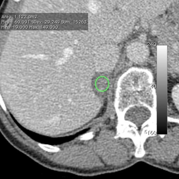
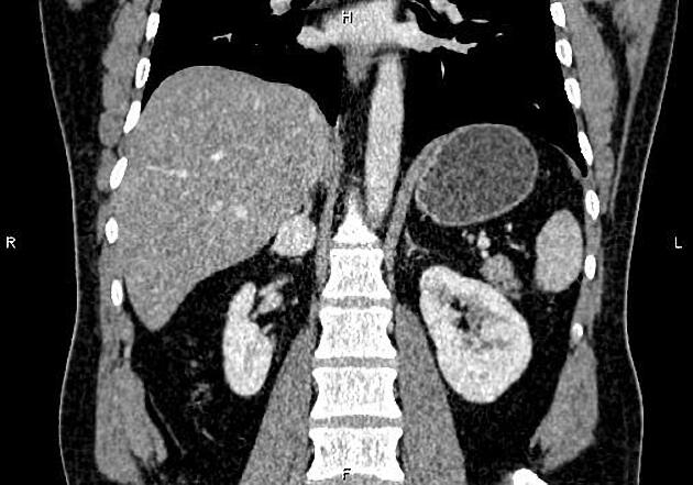
Adrenal washout can be calculated using the density value of an adrenal mass on non-enhanced, portal venous phase and 15-minute delayed CT scans (density measured in Hounsfield units (HU)). It is primarily used to diagnose adrenal adenoma.
-
absolute washout
-
relative washout
Practical points
adrenal washout has limited use when assessing incidental adrenal nodules <4 cm in size and >10 HU in patients without known malignancy 8
lesions that are inhomogeneous with large areas of necrosis or haemorrhage cannot be characterised by their washout pattern
phaeochromocytomas and hypervascular metastases (e.g. renal cell carcinoma, hepatocellular carcinoma) may also washout but should have a different clinical presentation; they also may have a higher absolute attenuation on the contrast phase (arterial or portal venous)
ADVERTISEMENT: Supporters see fewer/no ads
External links
If any of these links are broken or for other problems and questions, please contact editors@radiopaedia.org.
Which of the following adrenal gland lesions can potentially demonstrate rapid contrast washout on dynamic CT scans?
-
1. Johnson P, Horton K, Fishman E. Adrenal Mass Imaging with Multidetector CT: Pathologic Conditions, Pearls, and Pitfalls. Radiographics. 2009;29(5):1333-51. doi:10.1148/rg.295095027 - Pubmed
-
2. Patel J, Davenport M, Cohan R, Caoili E. Can Established CT Attenuation and Washout Criteria for Adrenal Adenoma Accurately Exclude Pheochromocytoma? AJR Am J Roentgenol. 2013;201(1):122-7. doi:10.2214/AJR.12.9620 - Pubmed
-
3. Blake M, Cronin C, Boland G. Adrenal Imaging. AJR Am J Roentgenol. 2010;194(6):1450-60. doi:10.2214/AJR.10.4547 - Pubmed
-
4. Blake M, Kalra M, Sweeney A et al. Distinguishing Benign from Malignant Adrenal Masses: Multi-Detector Row CT Protocol with 10-Minute Delay. Radiology. 2006;238(2):578-85. doi:10.1148/radiol.2382041514 - Pubmed
-
5. Northcutt B, Raman S, Long C et al. MDCT of Adrenal Masses: Can Dual-Phase Enhancement Patterns Be Used to Differentiate Adenoma and Pheochromocytoma? AJR Am J Roentgenol. 2013;201(4):834-9. doi:10.2214/AJR.12.9753 - Pubmed
-
6. Sahdev A, Willatt J, Francis I, Reznek R. The Indeterminate Adrenal Lesion. Cancer Imaging. 2010;10(1):102-13. doi:10.1102/1470-7330.2010.0012 - Pubmed
-
7. Nandra G, Duxbury O, Patel P, Patel J, Patel N, Vlahos I. Technical and Interpretive Pitfalls in Adrenal Imaging. Radiographics. 2020;40(4):1041-60. doi:10.1148/rg.2020190080 - Pubmed
-
8. Corwin M, Badawy M, Caoili E et al. Incidental Adrenal Nodules in Patients Without Known Malignancy: Prevalence of Malignancy and Utility of Washout CT for Characterization-A Multiinstitutional Study. AJR Am J Roentgenol. 2022;219(5):804-12. doi:10.2214/AJR.22.27901 - Pubmed
Multiple choice questions:
Promoted articles (advertising)


 Unable to process the form. Check for errors and try again.