
Presentation
Cushings syndrome.
Patient Data
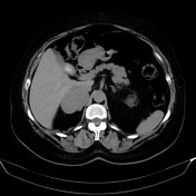

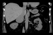
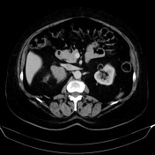
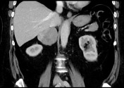
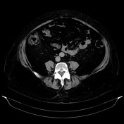
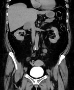

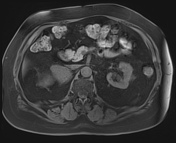
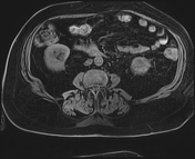
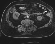
Large right adrenal mass lesion. This lesion is relatively well demarcated and abuts the posterior margin of the IVC, medial margin of the liver and lateral margin of right hemidiaphragm crura. There is no evidence of local invasion.
The more prominently enhancing posterosuperior component measures 29 HU on non-contrast, 84 HU on portal venous and 43 HU on delayed phase - calculating to 74% absolute washout and 49% relative washout.
The anterior component has less prominent enhancement and measures 35 HU on non-contrast, 59 HU on portal venous and 43 HU on delayed - calculating to 66% absolute washout and 27% relative washout.
Normal left adrenal gland. No hepatic mass. No lymphadenopathy.

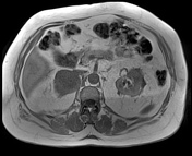

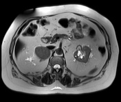
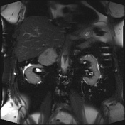
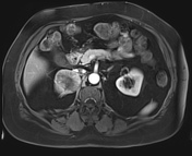
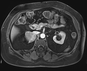
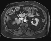
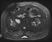
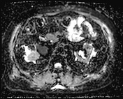
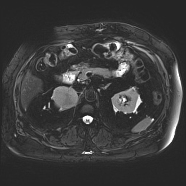
There is a large right adrenal mass that is bilobed in appearance. There is an anterosuperior focus of calcification or hemorrhage. There is prominent enhancement with band-like central stroma with enhancement also present on delayed imaging. The mass demonstrates abnormally restricted diffusion. No loss of signal on in and out-of-phase T1 imaging.
Case Discussion
The imaging features of this mass are not particularly suspicious for malignancy, although the size (>5 cm) and heterogeneity, along with the endocrine abnormality that prompted imaging, are worrisome.
Absolute washout is >60% in the posterior component, which is compatible with an adenoma; however, the anterior component has a relative washout of <40%, which is indeterminate.
MRI shows no fat signal drop-out, which is compatible with a lipid-poor adenoma; however, the restricted diffusion is concerning.
The patient proceeded to resection, which demonstrated an adrenal cortical carcinoma.


 Unable to process the form. Check for errors and try again.