
Apparent diffusion coefficient
Citation, DOI, disclosures and article data
At the time the article was created Mohammad Taghi Niknejad had no recorded disclosures.
View Mohammad Taghi Niknejad's current disclosuresAt the time the article was last revised Daniel J Bell had no financial relationships to ineligible companies to disclose.
View Daniel J Bell's current disclosures- ADC
- ADC map
- ADC maps
- ADC MRI
- Apparent diffusion coefficient
- Apparent diffusion coefficient map
- Apparent diffusion coefficient maps
- Apparent diffusion coefficient map (ADC map)
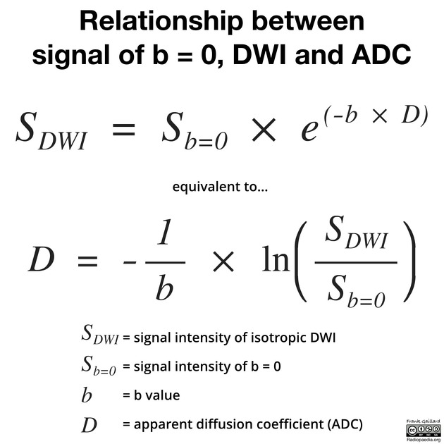
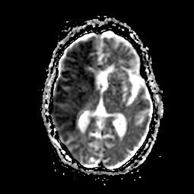
Apparent diffusion coefficient (ADC) is a measure of the magnitude of diffusion (of water molecules) within tissue, and is commonly clinically calculated using MRI with diffusion-weighted imaging (DWI) 1.
On this page:
Basics
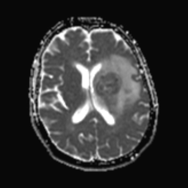
Diffusion-weighted imaging (DWI) is widely appreciated as an indispensable tool in the examination of the CNS. It is considered useful not only for the detection of acute ischemic stroke but also for the characterization and differentiation of brain tumors and intracranial infections.
DWI exploits the random motion of water molecules. The extent of tissue cellularity and the presence of intact cell membrane help determine the impedance of water molecule diffusion. This impedance of water molecules diffusion can be quantitatively assessed using the apparent diffusion coefficient (ADC) value. This assessment can be done using different b values via changing gradient amplitude 2,3,6.
Measurement
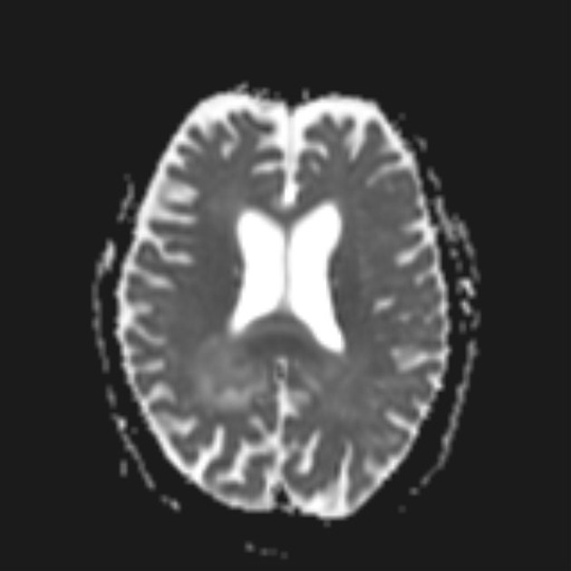
ADC values are calculated automatically by the software and then displayed as a parametric map that reflects the degree of diffusion of water molecules through different tissues. Then, by use of a dedicated workstation, ADC measurements are recorded for a given region by drawing regions of interest (ROIs) on the ADC map 6.
An ADC of tissue is expressed in units of mm2/s. There is no unanimity regarding the boundaries of the range of normal diffusion, but ADC values less than 1.0 to 1.1 x 10⁻³ mm2/s (or 1000-1100 x 10-6 mm2/s) are generally acknowledged in adults as indicating restriction. However, this is entirely dependent on the organ being imaged and the pathology 7.
Some rough useful values (10-6 mm2/s) 8-10:
-
brain
white matter: 670-800
deep grey matter: 700-850
cortical grey matter: 800-1000
CSF: 3000-3400
-
grade II: 1273 ± 293
grade III: 1067 ± 276
grade IV: 745 ± 135
-
pelvis 11,12
normal endometrial zone: 1530
-
benign endometrial lesions: 1300
uterine polyps: 1270-1580
endometrial cancer: 880-980
Diffusion changes are correlated to the clinical deficit and are potentially useful for early diagnosis and longitudinal evaluation, especially in the context of pharmacological trials.
See also
Quiz questions
References
- 1. Sener RN. Diffusion MRI: apparent diffusion coefficient (ADC) values in the normal brain and a classification of brain disorders based on ADC values. Comput Med Imaging Graph. 2001;25 (4): 299-326. Pubmed citation
- 2. Mascalchi M, Filippi M, Floris R et-al. Diffusion-weighted MR of the brain: methodology and clinical application. Radiol Med. 2005;109 (3): 155-97. Pubmed citation
- 3. Rana S, Albayram S, Lin DD et-al. Diffusion-weighted imaging and apparent diffusion coefficient maps in a case of intracerebral abscess with ventricular extension. AJNR Am J Neuroradiol. 2002;23 (1): 109-12. AJNR Am J Neuroradiol (full text) - Pubmed citation
- 4. Kang Y, Choi SH, Kim YJ et-al. Gliomas: Histogram analysis of apparent diffusion coefficient maps with standard- or high-b-value diffusion-weighted MR imaging-correlation with tumor grade. Radiology. 2011;261 (3): 882-90. Radiology (full text) - doi:10.1148/radiol.11110686 - Pubmed citation
- 5. . Magnetic Resonance Tomography. Springer. (2008) ISBN:3540293558. Read it at Google Books - Find it at Amazon
- 6. El Kady RM, Choudhary AK, Tappouni R. Accuracy of apparent diffusion coefficient value measurement on PACS workstation: A comparative analysis. AJR Am J Roentgenol. 2011;196 (3): W280-4. AJR Am J Roentgenol (full text) - doi:10.2214/AJR.10.4706 - Pubmed citation
- 7. Haaga JR, Boll D. CT and MRI of the whole body. Mosby. (2009) ISBN:0323053750. Read it at Google Books - Find it at Amazon
- 8. Helenius J, Soinne L, Perkiö J et-al. Diffusion-weighted MR imaging in normal human brains in various age groups. AJNR Am J Neuroradiol. 2002;23 (2): 194-9. AJNR Am J Neuroradiol (full text) - Pubmed citation
- 9. Annet L, Duprez T, Grandin C et-al. Apparent diffusion coefficient measurements within intracranial epidermoid cysts in six patients. Neuroradiology. 2002;44 (4): 326-8. doi:10.1007/s00234-001-0726-2 - Pubmed citation
- 10. Hilario A, Ramos A, Perez-Nuñez A et-al. The added value of apparent diffusion coefficient to cerebral blood volume in the preoperative grading of diffuse gliomas. AJNR Am J Neuroradiol. 2012;33 (4): 701-7. doi:10.3174/ajnr.A2846 - Pubmed citation
- 11. Tamai K, Koyama T, Saga T, Morisawa N, Fujimoto K, Mikami Y, Togashi K. The utility of diffusion-weighted MR imaging for differentiating uterine sarcomas from benign leiomyomas. (2008) European radiology. 18 (4): 723-30. doi:10.1007/s00330-007-0787-7 - Pubmed
- 12. Fujii S, Matsusue E, Kigawa J, Sato S, Kanasaki Y, Nakanishi J, Sugihara S, Kaminou T, Terakawa N, Ogawa T. Diagnostic accuracy of the apparent diffusion coefficient in differentiating benign from malignant uterine endometrial cavity lesions: initial results. (2008) European radiology. 18 (2): 384-9. doi:10.1007/s00330-007-0769-9 - Pubmed
Incoming Links
- Primary central nervous system posttransplant lymphoproliferative disorder
- Chronic pancreatitis
- Diffusion tensor imaging and fiber tractography
- T2 washout
- Cerebellar restricted diffusion
- Atypical meningioma
- Oesophageal MRI
- Prostate MRI (an approach)
- Clermont score (Crohn disease)
- Diffusion-weighted imaging
- MRI pulse sequence abbreviations
- Cerebral restricted diffusion
- Osseous Tumour Reporting and Data System (OT-RADS)
- Prostate Imaging-Reporting and Data System (PI-RADS)
- Quick stroke protocol (MRI)
- Physics curriculum
- Uterine restricted diffusion
- Supratentorial intracranial mass in an adult (an approach)
- Pineal and tectal plate protocol (MRI)
- Anal canal cancer protocol (MRI)
- Diffuse midline glioma
- Hypoglycaemic encephalopathy
- Atypical choroid plexus papilloma
- MCA infarction with subarachnoid FLAIR vascular hyperintensities
- Epidermoid cyst of the cerebellopontine cistern
- Acute necrotizing encephalitis of childhood
- Gastrointestinal stromal tumour (GIST)
- Severe urinary tract infection with complicating prostatic abscess
- Breast lymphoma (MRI)
- Tonsillar lymphoma
- Acute phase of hemiconvulsion-hemiplegia epilepsy syndrome
- Intracranial epidermoid cyst - cerebellopontine angle
Related articles: Imaging technology
- imaging technology
- imaging physics
- imaging in practice
-
x-rays
- x-ray physics
- x-ray in practice
- x-ray production
- x-ray tube
- filters
- automatic exposure control (AEC)
- beam collimators
- grids
- air gap technique
- cassette
- intensifying screen
- x-ray film
- image intensifier
- digital radiography
- digital image
- mammography
- x-ray artifacts
- radiation units
- radiation safety
- radiation detectors
- fluoroscopy
-
computed tomography (CT)
- CT physics
- CT in practice
- CT technology
- CT image reconstruction
- CT image quality
- CT dose
-
CT contrast media
-
iodinated contrast media
- agents
- water soluble
- water insoluble
- vicarious contrast material excretion
- iodinated contrast media adverse reactions
- agents
- non-iodinated contrast media
-
iodinated contrast media
-
CT artifacts
- patient-based artifacts
- physics-based artifacts
- hardware-based artifacts
- ring artifact
- tube arcing
- out of field artifact
- air bubble artifact
- helical and multichannel artifacts
- CT safety
- history of CT
-
MRI
- MRI physics
- MRI in practice
- MRI hardware
- signal processing
-
MRI pulse sequences (basics | abbreviations | parameters)
- T1 weighted image
- T2 weighted image
- proton density weighted image
- chemical exchange saturation transfer
- CSF flow studies
- diffusion weighted imaging (DWI)
- echo-planar pulse sequences
- fat-suppressed imaging sequences
- gradient echo sequences
- inversion recovery sequences
- metal artifact reduction sequence (MARS)
-
perfusion-weighted imaging
- techniques
- derived values
- saturation recovery sequences
- spin echo sequences
- spiral pulse sequences
- susceptibility-weighted imaging (SWI)
- T1 rho
- MR angiography (and venography)
-
MR spectroscopy (MRS)
- 2-hydroxyglutarate peak: resonates at 2.25 ppm
- alanine peak: resonates at 1.48 ppm
- choline peak: resonates at 3.2 ppm
- citrate peak: resonates at 2.6 ppm
- creatine peak: resonates at 3.0 ppm
- functional MRI (fMRI)
- gamma-aminobutyric acid (GABA) peak: resonates at 2.2-2.4 ppm
- glutamine-glutamate peak: resonates at 2.2-2.4 ppm
- Hunter's angle
- lactate peak: resonates at 1.3 ppm
- lipids peak: resonates at 1.3 ppm
- myoinositol peak: resonates at 3.5 ppm
- MR fingerprinting
- N-acetylaspartate (NAA) peak: resonates at 2.0 ppm
- propylene glycol peak: resonates at 1.13 ppm
-
MRI artifacts
- MRI hardware and room shielding
- MRI software
- patient and physiologic motion
- tissue heterogeneity and foreign bodies
- Fourier transform and Nyquist sampling theorem
- MRI contrast agents
- MRI safety
-
ultrasound
- ultrasound physics
-
transducers
- linear array
- convex array
- phased array
- frame averaging (frame persistence)
- ultrasound image resolution
- imaging modes and display
- pulse-echo imaging
- real-time imaging
-
Doppler imaging
- Doppler effect
- color Doppler
- power Doppler
- B flow
- color box
- Doppler angle
- pulse repetition frequency and scale
- wall filter
- color write priority
- packet size (dwell time)
- peak systolic velocity
- end-diastolic velocity
- resistive index
- pulsatility index
- Reynolds number
- panoramic imaging
- compound imaging
- harmonic imaging
- elastography
- scanning modes
- 2D ultrasound
- 3D ultrasound
- 4D ultrasound
- M-mode
-
ultrasound artifacts
- acoustic shadowing
- acoustic enhancement
- beam width artifact
- reverberation artifact
- ring down artifact
- mirror image artifact
- side lobe artifact
- speckle artifact
- speed displacement artifact
- refraction artifact
- multipath artifact
- anisotropy
- electrical interference artifact
- hardware-related artifacts
- Doppler artifacts
- aliasing
- tissue vibration
- spectral broadening
- blooming
- motion (flash) artifact
- twinkling artifact
- acoustic streaming
- biological effects of ultrasound
- history of ultrasound
-
nuclear medicine
- nuclear medicine physics
- detectors
- tissue to background ratio
-
radiopharmaceuticals
- fundamentals of radiopharmaceuticals
- radiopharmaceutical labeling
- radiopharmaceutical production
- nuclear reactor produced radionuclides
- cyclotron produced radionuclides
- radiation detection
- dosimetry
- specific agents
- carbon-11
- chromium-51
- fluorine agents
- gallium agents
- Ga-67 citrate
- Ga-68
- iodine agents
-
I-123
- I-123 iodide
- I-123 ioflupane (DaTSCAN)
- I-123 ortho-iodohippurate
- I-131
-
MIBG scans
- I-123 MIBG
- I-131 MIBG
-
I-123
- indium agents
- In-111 Octreoscan
- In-111 OncoScint
- In-111 Prostascint
- In-111 oxine labeled WBC
- krypton-81m
- nitrogen-13
- oxygen-15
- phosphorus-32
- selenium-75
-
technetium agents
- Tc-99m DMSA
- Tc-99m DTPA
- Tc-99m DTPA aerosol
- Tc-99m HMPAO
- Tc-99m HMPAO labeled WBC
- Tc-99m MAA
- Tc-99m MAG3
- Tc-99m MDP
- Tc-99m mercaptoacetyltriglycine
- Tc-99m pertechnetate
- Tc-99m labeled RBC
- Tc-99m sestamibi
- Tc-99m sulfur colloid
- Tc-99m sulfur colloid (oral)
- thallium-201 chloride
- xenon agents
- in vivo therapeutic agents
- pharmaceuticals used in nuclear medicine
-
emerging methods in medical imaging
- radiography
- phase-contrast imaging
- CT
- deep-learning reconstruction
- photon counting CT
- virtual non-contrast imaging
- ultrasound
- magnetomotive ultrasound (MMUS)
- superb microvascular imaging
- ultrafast Doppler imaging
- ultrasound localization microscopy
- MRI
- nuclear medicine
- total body PET system
- immuno-PET
- miscellaneous
- radiography


 Unable to process the form. Check for errors and try again.