
Presentation
Seizure.
Patient Data
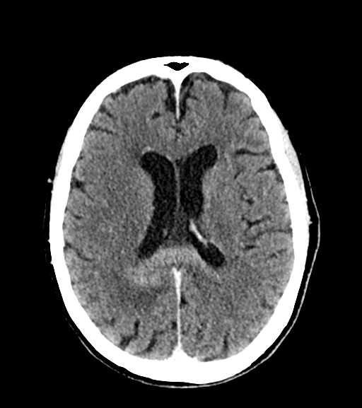
Enhancing intra-axial lesion crossing the splenium of the corpus callosum and additional enhancing mass centered in the fourth ventricle. Suggestion of rim of enhancement adjacent to the frontal horn right lateral ventricle.


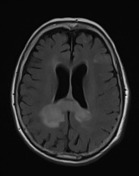
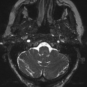
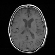

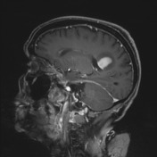
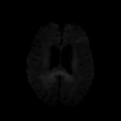

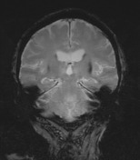
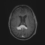
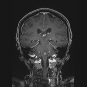
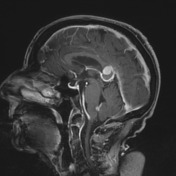
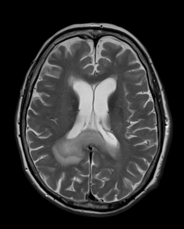
MRI shows vividly enhancing mass involving the splenium of the corpus callosum and subependymal extension within the anterior horns of the lateral ventricles and at the floor of the fourth ventricle. The corpus callosum prominent mass has diffusion restriction inferring high cellularity and surrounding vasogenic edema.
Case Discussion
Although not histologically proven, this case brings typical imaging features of CNS lymphoma characterized by a high-cellular periventricular mass with low ADC values and vivid and homogeneous contrast enhancement. Other differentials are felt unlikely.


 Unable to process the form. Check for errors and try again.