
Artery of Bernasconi and Cassinari
Citation, DOI, disclosures and article data
At the time the article was created Peter Fawzy had no recorded disclosures.
View Peter Fawzy's current disclosuresAt the time the article was last revised Henry Knipe had no recorded disclosures.
View Henry Knipe's current disclosures- Marginal tentorial artery
- Medial tentorial artery
- Medial tentorial artery of Bernasconi–Cassinari
- Recurrent artery of Bernasconi and Cassinari
- Artery of Bernasconi-Cassinari
- Marginal tentorial artery of Bernasconi–Cassinari
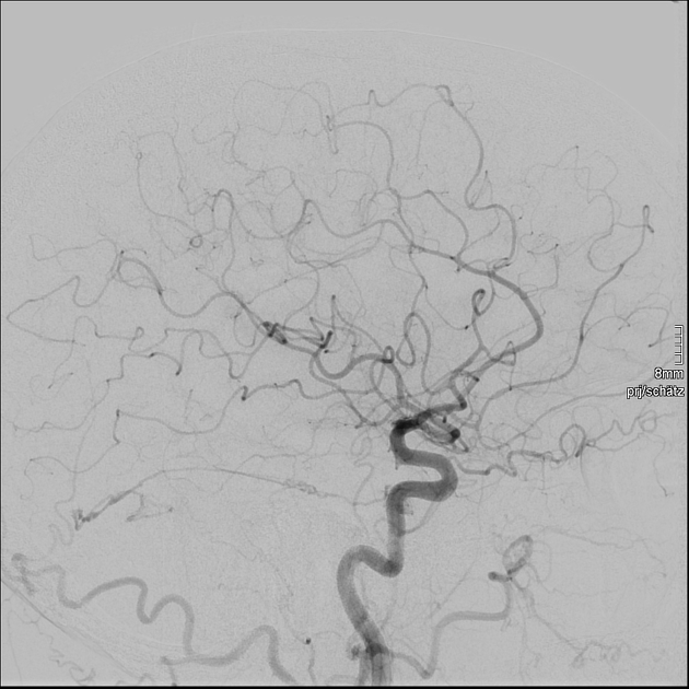
The artery of Bernasconi and Cassinari, also known as medial or marginal tentorial artery (of Bernasconi–Cassinari), commonly arises from the cavernous segment of the internal carotid artery.
On this page:
Gross anatomy
The artery of Bernasconi and Cassinari is ~2 cm long and is an important branch of the meningohypophyseal trunk, originating from the cavernous segment of the internal carotid artery and runs along the tentorium.
It is usually a single trunk, supplying the meninges of the tentorium cerebelli, and often supplies the oculomotor, trochlear, and abducens nerves.
Another branch of the meningohypophyseal trunk supplying the tentorium is the lateral tentorial artery (also known as the lateral tentorial arcade) which shows a course laterally towards the sigmoid sinus and on lateral projections it points in a downward direction.
Radiographic appearance
Due to its small size, the artery of Bernasconi and Cassinari is poorly visualised on angiography in the absence of pathologically increased blood flow, like in tentorial tumours and tentorial dural arteriovenous fistula (DAVF). When seen, it has a wavy appearance on angiography.
History and etymology
It was first described in 1957 by V Bernasconi and V Cassinari in the setting of tentorial meningiomas 3,4.
Related pathology
Lesions arising from the tentorium cerebelli, such as tentorial dural arteriovenous fistulas may derive arterial blood supply from the artery of Bernasconi and Cassinari.
The artery of Bernasconi and Cassinari is also the most likely artery to be enlarged on an angiogram in tentorial meningiomas.
Occasionally, it may provide essential collateral blood flow to the posterior circulation.
References
- 1. Liu S, Lee DC, Tanoura T. Tentorial dural arteriovenous fistula of the medial tentorial artery. (2016) Radiology case reports. 11 (3): 242-4. doi:10.1016/j.radcr.2016.06.006 - Pubmed
- 2. R. Shane Tubbs, Ha Son Nguyen, Mohammadali M. Shoja, Brion Benninger, Marios Loukas, Aaron A. Cohen-Gadol. The medial tentorial artery of Bernasconi–Cassinari: a comprehensive review of its anatomy and neurosurgical importance. (2011) Acta Neurochirurgica. 153 (12): 2485. doi:10.1007/s00701-011-1195-y - Pubmed
- 3. BERNASCONI V, CASSINARI V. [Angiographical characteristics of meningiomas of tentorium]. (1957) La Radiologia medica. 43 (10): 1015-26. Pubmed
- 4. A.D. Banerjee, H. Ezer, A. Nanda. The Artery of Bernasconi and Cassinari: A Morphometric Study for Superselective Catheterization. (2011) American Journal of Neuroradiology. 32 (9): 1751. doi:10.3174/ajnr.A2552 - Pubmed
Incoming Links
Related articles: Anatomy: Head and neck
- skeleton of the head and neck
-
cranial vault
- scalp (mnemonic)
- fontanelle
-
sutures
- calvarial
- facial
- frontozygomatic suture
- frontomaxillary suture
- frontolacrimal suture
- frontonasal suture
- temporozygomatic suture
- zygomaticomaxillary suture
- parietotemporal suture (parietomastoid suture)
- occipitotemporal suture (occipitomastoid suture)
- sphenofrontal suture
- sphenozygomatic suture
- spheno-occipital suture (not a true suture)
- lacrimomaxillary suture
- nasomaxillary suture
- internasal suture
- basal/internal
- skull landmarks
- frontal bone
- temporal bone
- parietal bone
- occipital bone
- skull base (foramina)
-
facial bones
- midline single bones
- paired bilateral bones
- cervical spine
- hyoid bone
- laryngeal cartilages
-
cranial vault
- muscles of the head and neck
- muscles of the tongue (mnemonic)
- muscles of mastication
-
facial muscles
- epicranius muscle
- circumorbital and palpebral muscles
- nasal muscles
-
buccolabial muscles
- elevators, retractors and evertors of the upper lip
- levator labii superioris alaeque nasalis muscle
- levator labii superioris muscle
- zygomaticus major muscle
- zygomaticus minor muscle
- levator anguli oris muscle
- malaris muscle
- risorius muscle
- depressors, retractors and evertors of the lower lip
- depressor labii inferioris muscle
- depressor anguli oris muscle
- mentalis muscle
- compound sphincter
-
orbicularis oris muscle
- incisivus labii superioris muscle
- incisivus labii inferioris muscle
-
orbicularis oris muscle
- muscle of mastication
- modiolus
- elevators, retractors and evertors of the upper lip
- muscles of the middle ear
- orbital muscles
- muscles of the soft palate
- pharyngeal muscles
- suprahyoid muscles
- infrahyoid muscles
- intrinsic muscles of the larynx
- muscles of the neck
- platysma muscle
- longus colli muscle
- longus capitis muscle
- scalenus anterior muscle
- scalenus medius muscle
- scalenus posterior muscle
- scalenus pleuralis muscle
- sternocleidomastoid muscle
-
suboccipital muscles
- rectus capitis posterior major muscle
- rectus capitis posterior minor muscle
- obliquus capitis superior muscle
- obliquus capitis inferior muscle
- accessory muscles of the neck
- deep cervical fascia
-
deep spaces of the neck
- anterior cervical space
- buccal space
- carotid space
- danger space
- deep cervical fascia
- infratemporal fossa
- masticator space
- parapharyngeal space
- stylomandibular tunnel
- parotid space
- pharyngeal (superficial) mucosal space
- perivertebral space
- posterior cervical space
- pterygopalatine fossa
- retropharyngeal space
- suprasternal space (of Burns)
- visceral space
- surgical triangles of the neck
- orbit
- ear
- paranasal sinuses
- upper respiratory tract
- viscera of the neck
- blood supply of the head and neck
-
arterial supply
-
common carotid artery
- carotid body
- carotid bifurcation
- subclavian artery
- variants
-
common carotid artery
- venous drainage
-
arterial supply
- innervation of the head and neck
-
cranial nerves
- olfactory nerve (CN I)
- optic nerve (CN II)
- oculomotor nerve (CN III)
- trochlear nerve (CN IV)
-
trigeminal nerve (CN V) (mnemonic)
- trigeminal ganglion
- ophthalmic division
- maxillary division
- mandibular division
- abducens nerve (CN VI)
- facial nerve (CN VII)
-
vestibulocochlear nerve (CN VIII)
- vestibular ganglion (Scarpa's ganglion)
- glossopharyngeal nerve (CN IX)
- vagus nerve (CN X)
- (spinal) accessory nerve (CN XI)
- hypoglossal nerve (CN XII)
- parasympathetic ganglia of the head and neck
- cervical sympathetic ganglia
- greater occipital nerve
- third occipital nerve
-
cervical plexus
- muscular branches
- longus capitis
- longus colli
- scalenes
- geniohyoid
- thyrohyoid
-
ansa cervicalis
- omohyoid (superior and inferior bellies separately)
- sternothyroid
- sternohyoid
- phrenic nerve
- contribution to the accessory nerve (CN XI)
- cutaneous branches
- muscular branches
- brachial plexus
- pharyngeal plexus
-
cranial nerves
- lymphatic drainage of the head and neck
- embryological development of the head and neck


 Unable to process the form. Check for errors and try again.