
Auriculotemporal nerve
Citation, DOI, disclosures and article data
At the time the article was created Craig Hacking had no recorded disclosures.
View Craig Hacking's current disclosuresAt the time the article was last revised Yoshi Yu had no financial relationships to ineligible companies to disclose.
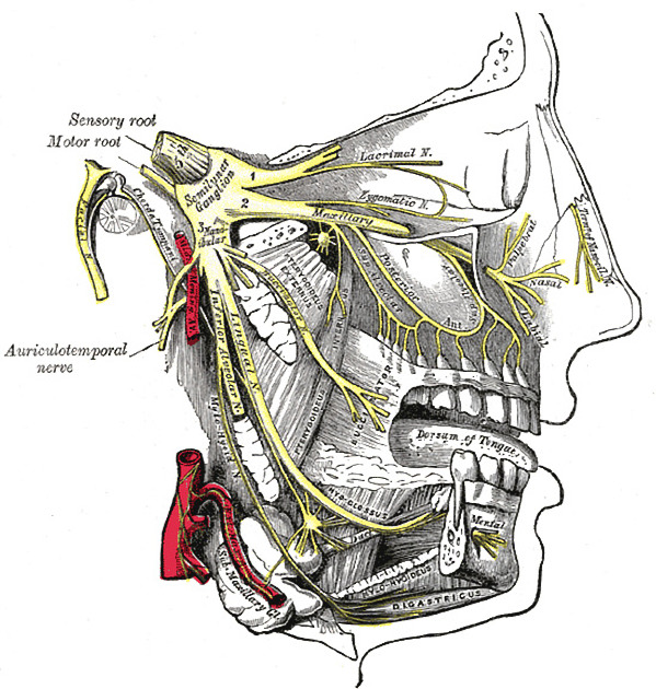
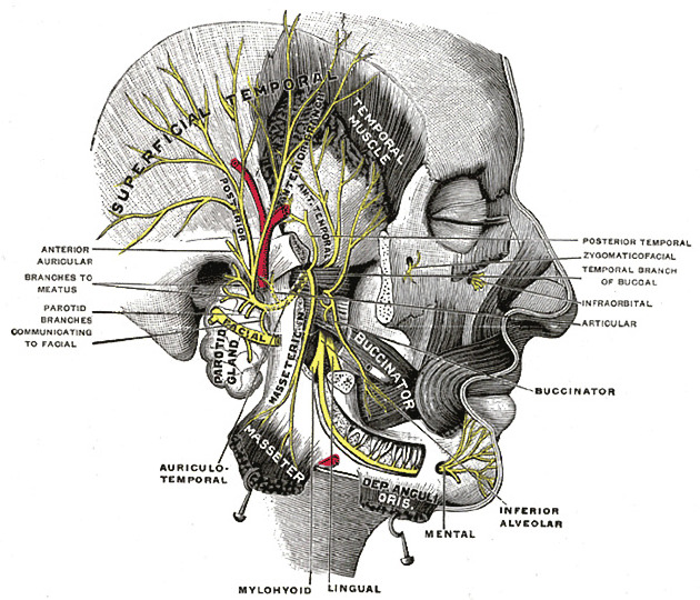
View Yoshi Yu's current disclosuresThe auriculotemporal nerve is a sensory branch of the posterior division of the mandibular division of the trigeminal nerve.
On this page:
Gross anatomy
The auriculotemporal nerve arises posteriorly from the posterior division of the mandibular nerve. It arises as two separate roots which encircle the middle meningeal artery as it ascends to enter the foramen spinosum. The two roots then rejoin to pass medial to the neck of the mandible, deep to the sphenomandibular ligament before ascending in between the temporomandibular joint and the external auditory meatus, giving off small articular branches to the temporomandibular joint. At the upper border of the parotid gland it supplies the sheath of the gland and then terminates as two cutaneous branches 1-3:
-
an auricular branch which supplies the:
external surface of the tympanic membrane along with the greater auricular nerve (internal surface is supplied by the tympanic branch of the glossopharyngeal nerve)
skin of the auricle anterior to the external acoustic meatus. The remainder of the auricle is supplied by the greater auricular nerve (C2, C3)
a temporal branch which ascends posterior to the superficial temporal vessels over the root of the zygoma to supply the posterior half of the temple, that is the ‘hairy skin’ of the temple; the anterior half of the temple ("skin of the hairless temple” is supplied by the zygomaticotemporal nerve from the maxillary division of the trigeminal nerve)
The auriculotemporal nerve receives sympathetic fibres from the middle meningeal artery and secretomotor parasympathetic fibres (which derive from the otic ganglion) from the lesser petrosal nerve, both of which supply the parotid gland 1-3.
There are communicating rami between the auriculotemporal nerve and the facial nerve within the parotid gland 4.
ADVERTISEMENT: Supporters see fewer/no ads
Radiographic features
The auriculotemporal nerve is usually not seen on CT or MRI. However, the expected course of the auriculotemporal nerve lies parallel to the posterior border of the mandible, just superior to the level of the terminal bifurcation of the external carotid artery into the maxillary and superficial temporal arteries 4.
Related pathology
perineural spread of head and neck malignancies (e.g. cutaneous squamous cell carcinoma, salivary gland adenoid cystic carcinoma) can occur along the auriculotemporal nerve, with potential to spread to the facial nerve from the mandibular division (V3) of the trigeminal nerve, or vice-versa 4.
Frey syndrome: commonest post-parotidectomy complication
Quiz questions
References
- 1. Keith L. Moore, Arthur F. Dalley. Clinically Oriented Anatomy. ISBN: 9780683061413
- 2. Robert H. Whitaker, Neil R. Borley. Instant Anatomy. ISBN: 9780632054039
- 3. Mcminn. Last's Anatomy. ISBN: 9780729537520
- 4. Schmalfuss IM, Tart RP, Mukherji S, Mancuso AA. Perineural tumor spread along the auriculotemporal nerve. (2002) AJNR. American journal of neuroradiology. 23 (2): 303-11. Pubmed
Incoming Links
- Antihelix (ear)
- Helix (ear)
- Greater wing of sphenoid
- Tympanic membrane
- Otic ganglion
- Trigeminal nerve
- Tragus
- Scalp nerve supply (mnemonic)
- External ear
- Sphenomandibular ligament
- Posterior division of the mandibular nerve
- Scalp
- Glossopharyngeal nerve
- Temporal fossa
- Frey syndrome
- Temporomandibular joint
- Parotid gland
- Zygomaticotemporal nerve
- Masseteric nerve
- Parasympathetic nervous system
Related articles: Anatomy: Head and neck
- skeleton of the head and neck[+][+]
-
cranial vault
- scalp (mnemonic)
- fontanelle
-
sutures
- calvarial
- facial
- frontozygomatic suture
- frontomaxillary suture
- frontolacrimal suture
- frontonasal suture
- temporozygomatic suture
- zygomaticomaxillary suture
- parietotemporal suture (parietomastoid suture)
- occipitotemporal suture (occipitomastoid suture)
- sphenofrontal suture
- sphenozygomatic suture
- spheno-occipital suture (not a true suture)
- lacrimomaxillary suture
- nasomaxillary suture
- internasal suture
- basal/internal
- skull landmarks
- frontal bone
- temporal bone
- parietal bone
- occipital bone
- skull base (foramina)
-
facial bones
- midline single bones
- paired bilateral bones
- cervical spine
- hyoid bone
- laryngeal cartilages
-
cranial vault
- muscles of the head and neck[+][+]
- muscles of the tongue (mnemonic)
- muscles of mastication
-
facial muscles
- epicranius muscle
- circumorbital and palpebral muscles
- nasal muscles
-
buccolabial muscles
- elevators, retractors and evertors of the upper lip
- levator labii superioris alaeque nasalis muscle
- levator labii superioris muscle
- zygomaticus major muscle
- zygomaticus minor muscle
- levator anguli oris muscle
- malaris muscle
- risorius muscle
- depressors, retractors and evertors of the lower lip
- depressor labii inferioris muscle
- depressor anguli oris muscle
- mentalis muscle
- compound sphincter
-
orbicularis oris muscle
- incisivus labii superioris muscle
- incisivus labii inferioris muscle
-
orbicularis oris muscle
- muscle of mastication
- modiolus
- elevators, retractors and evertors of the upper lip
- muscles of the middle ear
- orbital muscles
- muscles of the soft palate
- pharyngeal muscles
- suprahyoid muscles
- infrahyoid muscles
- intrinsic muscles of the larynx
- muscles of the neck
- platysma muscle
- longus colli muscle
- longus capitis muscle
- scalenus anterior muscle
- scalenus medius muscle
- scalenus posterior muscle
- scalenus pleuralis muscle
- sternocleidomastoid muscle
-
suboccipital muscles
- rectus capitis posterior major muscle
- rectus capitis posterior minor muscle
- obliquus capitis superior muscle
- obliquus capitis inferior muscle
- accessory muscles of the neck
- deep cervical fascia[+][+]
-
deep spaces of the neck[+][+]
- anterior cervical space
- buccal space
- carotid space
- danger space
- deep cervical fascia
- infratemporal fossa
- masticator space
- parapharyngeal space
- stylomandibular tunnel
- parotid space
- pharyngeal (superficial) mucosal space
- perivertebral space
- posterior cervical space
- pterygopalatine fossa
- retropharyngeal space
- suprasternal space (of Burns)
- visceral space
- surgical triangles of the neck[+][+]
- orbit[+][+]
- ear[+][+]
- paranasal sinuses[+][+]
- upper respiratory tract[+][+]
- viscera of the neck[+][+]
- blood supply of the head and neck[+][+]
-
arterial supply
-
common carotid artery
- carotid body
- carotid bifurcation
- subclavian artery
- variants
-
common carotid artery
- venous drainage
-
arterial supply
- innervation of the head and neck
-
cranial nerves
- olfactory nerve (CN I)
- optic nerve (CN II)
- oculomotor nerve (CN III)
- trochlear nerve (CN IV)
-
trigeminal nerve (CN V) (mnemonic)
- trigeminal ganglion
- ophthalmic division[+][+]
- maxillary division[+][+]
- mandibular division
- abducens nerve (CN VI)
- facial nerve (CN VII)[+][+]
-
vestibulocochlear nerve (CN VIII)[+][+]
- vestibular ganglion (Scarpa's ganglion)
- glossopharyngeal nerve (CN IX)[+][+]
- vagus nerve (CN X)[+][+]
- (spinal) accessory nerve (CN XI)
- hypoglossal nerve (CN XII)
- parasympathetic ganglia of the head and neck[+][+]
- cervical sympathetic ganglia[+][+]
- greater occipital nerve
- third occipital nerve
-
cervical plexus[+][+]
- muscular branches
- longus capitis
- longus colli
- scalenes
- geniohyoid
- thyrohyoid
-
ansa cervicalis
- omohyoid (superior and inferior bellies separately)
- sternothyroid
- sternohyoid
- phrenic nerve
- contribution to the accessory nerve (CN XI)
- cutaneous branches
- muscular branches
- brachial plexus
- pharyngeal plexus
-
cranial nerves
- lymphatic drainage of the head and neck[+][+]
- embryological development of the head and neck[+][+]


 Unable to process the form. Check for errors and try again.