
Bladder exstrophy (also known as ectopia vesicae) refers to a herniation of the urinary bladder through an anterior abdominal wall defect. The severity of these defects is widely variable.
On this page:
Epidemiology
The estimated incidence of bladder exstrophy is 1:10,000-50,000 live births 3,5. There is a recognised male predilection with a male-to-female ratio of ~3:1 6. Most cases are sporadic.
Pathology
Bladder exstrophy is thought to be caused by a developmental defect of the cloacal membrane, resulting in a subsequent eversion of the bladder mucosa. This then protrudes out as a mass-like lesion.
Associations
General
extension of the bladder defect into the urethra
bilateral inguinal herniation
OEIS complex (omphalocele, exstrophy of the cloaca, imperforate anus, and spinal defects)
In females
Serological markers
raised maternal alpha-fetoprotein levels
Radiographic features
Imaging findings include a soft-tissue mass extending from a large infra-umbilical anterior wall defect which may be close to the umbilical arterial exits. The absence of a normal urinary bladder and a low-lying umbilical cord insertion 4 may also indicate the diagnosis.
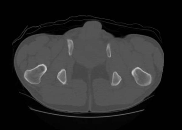
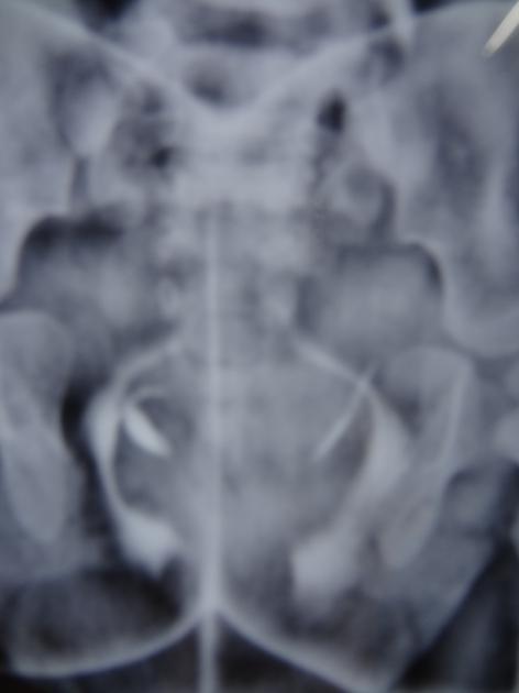
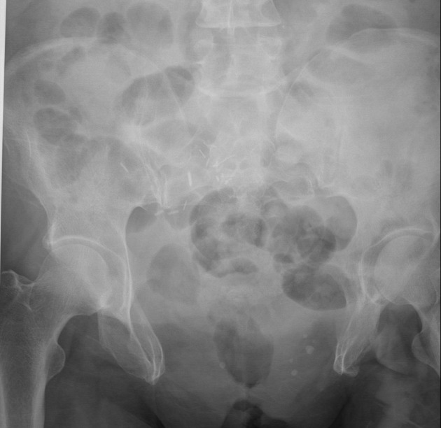
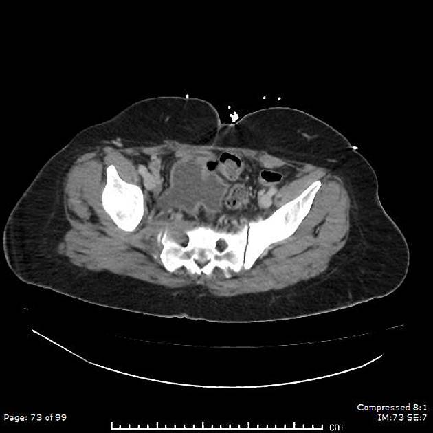
Failure of the pubic bones to meet in the midline (widened pubic symphysis). There will be shortening of the pubic rami and acetabular retroversion. This appearance on AP plain radiograph of the pelvis has been likened to a manta ray swimming towards you (manta ray sign) 7. Hurley stick appearance of distal ureters has been described in excretory urogram 8.
Amniotic fluid volumes are often normal.
Treatment and prognosis
Treatment is with surgical intervention - staged multidisciplinary reconstruction and the prognosis is generally good.
Complications
increased incidence of malignancy in the extruded bladder
See also
cloacal exstrophy: more severe anomaly


 Unable to process the form. Check for errors and try again.