
Carcinoid tumours of the lung
Citation, DOI, disclosures and article data
At the time the article was created Yuranga Weerakkody had no recorded disclosures.
View Yuranga Weerakkody's current disclosuresAt the time the article was last revised Henry Knipe had the following disclosures:
- Integral Diagnostics, Shareholder (ongoing)
- Micro-X Ltd, Shareholder (ongoing)
These were assessed during peer review and were determined to not be relevant to the changes that were made.
View Henry Knipe's current disclosures- Pulmonary carcinoid tumours
- Pulmonary carcinoids
- Carcinoid tumours of lung
- Carcinoid tumour of the lung
- Pulmonary carcinoid tumors
- Carcinoid tumour of lung
- Carcinoid tumor of the lung
- Carcinoid tumor of lung
- Pulmonary carcinoid tumour
- Pulmonary carcinoid tumor
- Pulmonary carcinoid
Carcinoid tumours of the lung are a subgroup of neuroendocrine tumours of the lung, of lower grade than small cell carcinoma of the lung and large cell neuroendocrine carcinoma of the lung.
For a general discussion, please refer to the article on carcinoid tumours.
Pathology
Carcinoid tumours can be divided into two groups dependent on location:
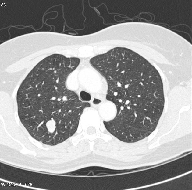
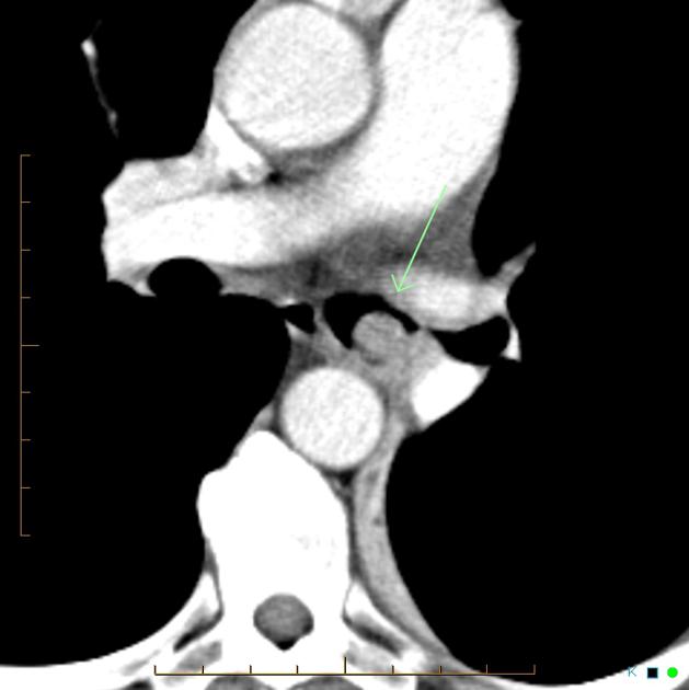
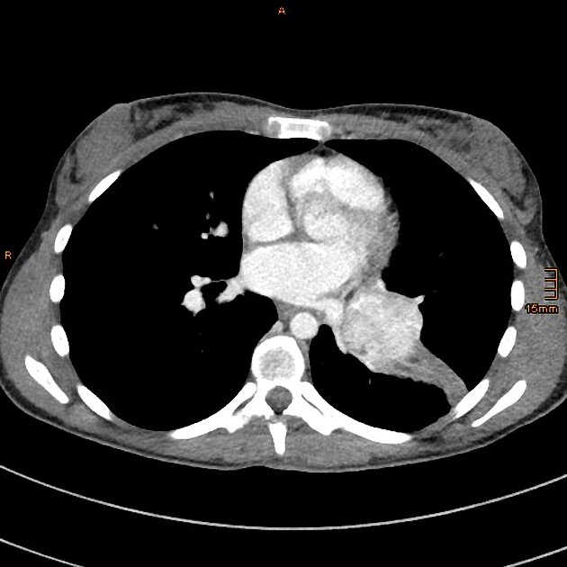
bronchial carcinoid tumours: central lesions (most common ~75% 9)
peripheral pulmonary carcinoid tumours: peripheral lesions (less common reported range 16-40% 9)
Carcinoid tumours can also be divided into two histological groups (requires resected specimen rather than biopsy 6) as follows:
-
typical carcinoid tumours of the lung
considered much more common (~90%) 5,9
low grade/well-differentiated 4
-
atypical carcinoid tumours of the lung
less well-differentiated
more aggressive 3,4
Radiographic features
Please refer to location-dependent subtypes above.
References
- 1. Chong S, Lee K, Chung M, Han J, Kwon O, Kim T. Neuroendocrine Tumors of the Lung: Clinical, Pathologic, and Imaging Findings. Radiographics. 2006;26(1):41-57; discussion 57. doi:10.1148/rg.261055057 - Pubmed
- 2. Magid D, Siegelman S, Eggleston J, Fishman E, Zerhouni E. Pulmonary Carcinoid Tumors: CT Assessment. J Comput Assist Tomogr. 1989;13(2):244-7. doi:10.1097/00004728-198903000-00010 - Pubmed
- 3. Marty-Ané C, Costes V, Pujol J, Alauzen M, Baldet P, Mary H. Carcinoid Tumors of the Lung: Do Atypical Features Require Aggressive Management? Ann Thorac Surg. 1995;59(1):78-83. doi:10.1016/0003-4975(94)00630-P - Pubmed
- 4. Thomas C, Tazelaar H, Jett J. Typical and Atypical Pulmonary Carcinoids : Outcome in Patients Presenting with Regional Lymph Node Involvement. Chest. 2001;119(4):1143-50. doi:10.1378/chest.119.4.1143 - Pubmed
- 5. Cooper W, Thourani V, Gal A, Lee R, Mansour K, Miller J. The Surgical Spectrum of Pulmonary Neuroendocrine Neoplasms. Chest. 2001;119(1):14-8. doi:10.1378/chest.119.1.14 - Pubmed
- 6. Rekhtman N. Neuroendocrine Tumors of the Lung: An Update. Arch Pathol Lab Med. 2010;134(11):1628-38. doi:10.5858/2009-0583-RAR.1 - Pubmed
- 7. Tsubochi H, Endo S, Oda Y, Dobashi Y. Carcinoid Tumor of the Lung with Massive Ossification: Report of a Case Showing the Evidence of Osteomimicry and Review of the Literature. Int J Clin Exp Pathol. 2013;6(5):957-61. PMC3638109 - Pubmed
- 8. Jeung M, Gasser B, Gangi A et al. Bronchial Carcinoid Tumors of the Thorax: Spectrum of Radiologic Findings. Radiographics. 2002;22(2):351-65. doi:10.1148/radiographics.22.2.g02mr01351 - Pubmed
- 9. Papaporfyriou A, Domayer J, Meilinger M et al. Bronchoscopic Diagnosis and Treatment of Endobronchial Carcinoid: Case Report and Review of the Literature. Eur Respir Rev. 2021;30(159):200115. doi:10.1183/16000617.0115-2020 - Pubmed
Incoming Links
- Pulmonary mass
- Solitary pulmonary nodule
- Peripheral, pleura-based and perifissural nodules
- Non-small cell lung cancer
- Endobronchial metastases (mnemonic)
- Peripheral pulmonary carcinoid tumour
- Well-differentiated neuroendocrine tumour of the prostate
- Atypical pulmonary carcinoid tumour
- Benign vs malignant pulmonary nodule
- Bronchial atresia
- Calcified pulmonary nodules
- Pulmonary neuroendocrine tumours
- Pulmonary tumourlet
- Hyperattenuating pulmonary mass lesion
- Unilateral hypertransradiant hemithorax
- MIBG
- Von Hippel-Lindau disease
- Carcinoid tumour of the lung
- Carcinoid tumor of the lung
- Typical pulmonary carcinoid tumor
- Carcinoid tumor causing complete atelectasis left lung
- Bronchial carcinoid tumour
- Bronchial carcinoid tumour causing right lower lobe collapse
- Bronchial carcinoid
- Bronchial carcinoid tumor
- Endobronchial carcinoid tumour
- Diffuse idiopathic pulmonary neuroendocrine cell hyperplasia with pulmonary carcinoid tumour
- Bronchial carcinoid tumor
- Bronchus intermedius carcinoid tumour
- Obstructing typical carcinoid causing bronchoceles
- Pulmonary carcinoid
- Bronchial carcinoid tumour with right lower lobe collapse
- Pulmonary carcinoid tumour - peripheral
Related articles: Chest
- imaging techniques
-
chest radiograph
- radiography
-
approach
- ABCDE
- ABCDEFGHI
- congenital heart disease
- medical devices in the thorax
- common lines and tubes
- nasogastric tubes
- endotracheal tubes
- central venous catheters
- oesophageal temperature probe
- tracheostomy tube
- pleural catheters
- cardiac conduction devices
- prosthetic heart valve
- review areas
-
airspace opacification
- differential diagnoses of airspace opacification
- lobar consolidation
-
atelectasis
- mechanism-based
- morphology-based
- lobar lung collapse
- chest x-ray in the exam setting
- cardiomediastinal contour
- chest radiograph zones
- tracheal air column
- fissures
- normal chest x-ray appearance of the diaphragm
- nipple shadow
-
lines and stripes
- anterior junction line
- posterior junction line
- right paratracheal stripe
- left paratracheal stripe
- posterior tracheal stripe/tracheo-oesophageal stripe
- posterior wall of bronchus intermedius
- right paraspinal line
- left paraspinal line
- aortic-pulmonary stripe
- aortopulmonary window
- azygo-oesophageal recess
- spaces
- signs
- air bronchogram
- big rib sign
- Chang sign
- Chen sign
- coin lesion
- continuous diaphragm sign
- dense hilum sign
- double contour sign
- egg-on-a-string sign
- extrapleural sign
- finger in glove sign
- flat waist sign
- Fleischner sign
- ginkgo leaf sign
- Golden S sign
- Hampton hump
- haystack sign
- hilum convergence sign
- hilum overlay sign
- Hoffman-Rigler sign
- holly leaf sign
- incomplete border sign
- juxtaphrenic peak sign
- Kirklin sign
- medial stripe sign
- melting ice cube sign
- more black sign
- Naclerio V sign
- Palla sign
- pericardial fat tag sign
- Shmoo sign
- silhouette sign
- snowman sign
- spinnaker sign
- steeple sign
- straight left heart border sign
- third mogul sign
- tram-track sign
- walking man sign
- water bottle sign
- wave sign
- Westermark sign
- HRCT
-
chest radiograph
- airways
- bronchitis
- small airways disease
-
bronchiectasis
- broncho-arterial ratio
- related conditions
- differentials by distribution
- narrowing
-
tracheal stenosis
- diffuse tracheal narrowing (differential)
-
bronchial stenosis
- diffuse airway narrowing (differential)
-
tracheal stenosis
- diverticula
- pulmonary oedema
-
interstitial lung disease (ILD)
- Anti-Jo-1 antibody-positive interstitial lung disease
- drug-induced interstitial lung disease
-
hypersensitivity pneumonitis
- acute hypersensitivity pneumonitis
- subacute hypersensitivity pneumonitis
- chronic hypersensitivity pneumonitis
- aetiology
- bird fancier's lung: pigeon fancier's lung
- farmer's lung
- cheese workers' lung
- bagassosis
- mushroom worker’s lung
- malt worker’s lung
- maple bark disease
- hot tub lung
- wine maker’s lung
- woodsman’s disease
- thatched roof lung
- tobacco grower’s lung
- potato riddler’s lung
- summer-type pneumonitis
- dry rot lung
- machine operator’s lung
- humidifier lung
- shower curtain disease
- furrier’s lung
- miller’s lung
- lycoperdonosis
- saxophone lung
-
idiopathic interstitial pneumonia (mnemonic)
- acute interstitial pneumonia (AIP)
- cryptogenic organising pneumonia (COP)
- desquamative interstitial pneumonia (DIP)
- non-specific interstitial pneumonia (NSIP)
- idiopathic pleuroparenchymal fibroelastosis
- lymphoid interstitial pneumonia (LIP)
- respiratory bronchiolitis–associated interstitial lung disease (RB-ILD)
- usual interstitial pneumonia / idiopathic pulmonary fibrosis (UIP/IPF)
-
pneumoconioses
- fibrotic
- non-fibrotic
-
lung cancer
-
non-small-cell lung cancer
-
adenocarcinoma
- pre-invasive tumours
- minimally invasive tumours
- invasive tumours
- variants of invasive carcinoma
- described imaging features
- adenosquamous carcinoma
- large cell carcinoma
- primary sarcomatoid carcinoma of the lung
- squamous cell carcinoma
- salivary gland-type tumours
-
adenocarcinoma
- pulmonary neuroendocrine tumours
- preinvasive lesions
-
lung cancer invasion patterns
- tumour spread through air spaces (STAS)
- presence of non-lepidic patterns such as acinar, papillary, solid, or micropapillary
- myofibroblastic stroma associated with invasive tumour cells
- pleural invasion
- vascular invasion
- tumours by location
- benign neoplasms
- pulmonary metastases
- lung cancer screening
- lung cancer staging
-
non-small-cell lung cancer


 Unable to process the form. Check for errors and try again.