
Presentation
Incidental finding on routine chest radiograph.
Patient Data
Gender: Female

Download
Info
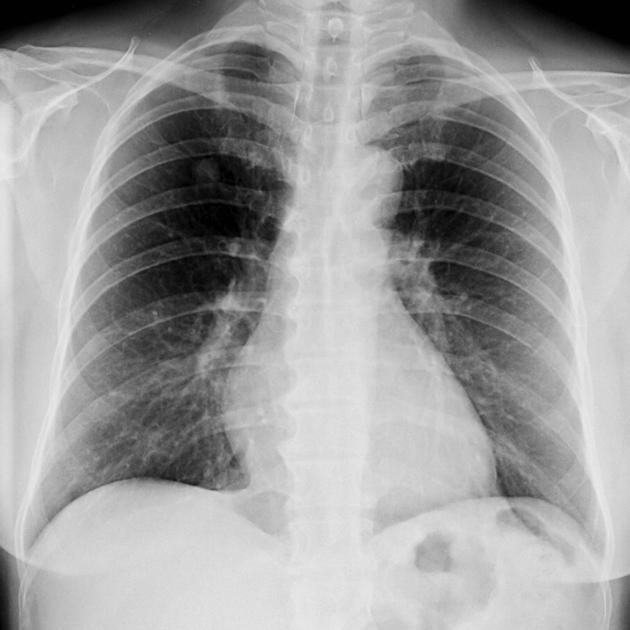
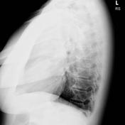
Circumscribed lung nodule in the right upper zone. This is confirmed as being within the right upper lobe on lateral radiograph.


Download
Info
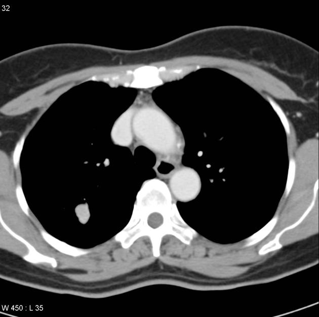
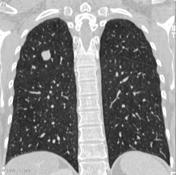
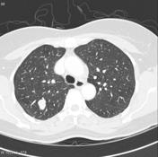
A 1.5 cm lung nodule in the right upper lobe with lobulated margin. A subsegmental bronchiole is seen leading to the nodule. The nodule mildly enhances and is concerning for malignancy.
Case Discussion
The differential is wide and includes primary lung malignancy, metastatic disease and benign tumor.
This patient underwent a right upper lobectomy. Histology confirmed typical carcinoid tumor of the lung, with two further smaller tumourlets within the right upper lobe which were not appreciated on CT.


 Unable to process the form. Check for errors and try again.