
Carotid cave
Citation, DOI, disclosures and article data
At the time the article was created Ayush Goel had no recorded disclosures.
View Ayush Goel's current disclosuresAt the time the article was last revised P. Vinícius Staziaki had no recorded disclosures.
View P. Vinícius Staziaki's current disclosures- Carotid caves
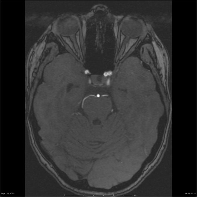
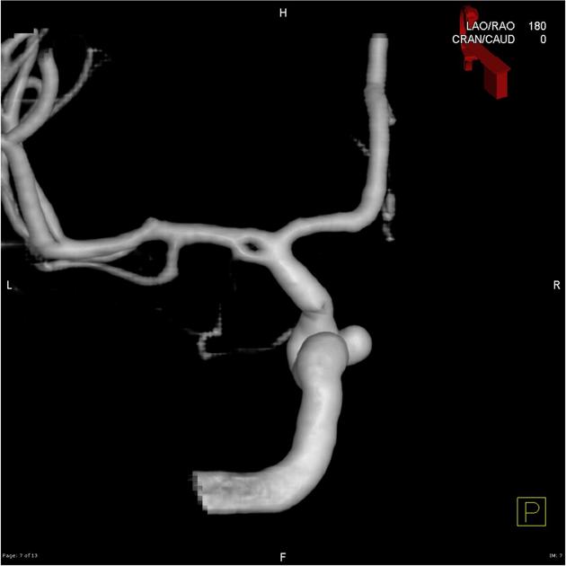
The carotid cave is a potential dural space formed by the redundant distal dural ring on the medial aspect of the clinoid segment of the internal carotid artery (ICA). It has been reported to be present in ~80% of cadaveric specimens 3.
On this page:
Gross anatomy
The clinoid segment of the ICA is bounded by proximal and distal dural rings that are variably adherent to each other on the anterior aspect. The distal dural ring is inclined posteromedially, tightly adherent to the lateral aspect of ICA, but loosely adherent to the medial aspect - creating the potential space that is the carotid cave.
Related pathology
The clinical significance of this region lies in the fact that aneurysms projecting medially from the ICA in this location (commonly referred to as carotid cave aneurysms) may be entirely extradural or may have an intradural component.
If entirely extradural in location, rupture does not produce subarachnoid haemorrhage (SAH). On the other hand, if partially intradural (typically at the neck), then subarachnoid haemorrhage is a possibility, and thus treatment potentially required.
Radiographic features
On imaging (CT, MRI, DSA) the relationship of the aneurysm to the distal dural ring can be difficult to establish. As such, these are called transitional aneurysms, with potential for SAH.
See also
References
- 1. Joo W, Funaki T, Yoshioka F et-al. Microsurgical anatomy of the carotid cave. Neurosurgery. 2012;70 (2 Suppl Operative): 300-11. doi:10.1227/NEU.0b013e3182431767 - Pubmed citation
- 2. Kobayashi S, Kyoshima K, Gibo H et-al. Carotid cave aneurysms of the internal carotid artery. J. Neurosurg. 1989;70 (2): 216-21. doi:10.3171/jns.1989.70.2.0216 - Pubmed citation
- 3. Lee N, Jung JY, Huh SK et-al. Distinction between Intradural and Extradural Aneurysms Involving the Paraclinoid Internal Carotid Artery with T2-Weighted Three-Dimensional Fast Spin-Echo Magnetic Resonance Imaging. J Korean Neurosurg Soc. 2010;47 (6): 437-41. doi:10.3340/jkns.2010.47.6.437 - Free text at pubmed - Pubmed citation
Incoming Links
Related articles: Anatomy: Brain
-
brain
- grey matter
- white matter
-
cerebrum
-
cerebral hemisphere (telencephalon)
- cerebral lobes and gyri
- frontal lobe
- parietal lobe
-
occipital lobe
- occipital pole
- lingual gyrus
- fusiform gyrus (Brodmann area 37)
- calcarine (visual) cortex
- cuneus
- temporal lobe
- basal forebrain
- limbic system
- insula
-
cerebral sulci and fissures (A-Z)
- calcarine fissure
- callosal sulcus
- central (Rolandic) sulcus
- cingulate sulcus
- collateral sulcus
- inferior frontal sulcus
- inferior occipital sulcus
- inferior temporal sulcus
- interhemispheric fissure
- intraparietal sulcus
- lateral (Sylvian) sulcus
- lateral occipital sulcus
- marginal sulcus
- occipitotemporal sulcus
- olfactory sulcus
- paracentral sulcus
- paraolfactory sulcus
- parieto-occipital fissure
- posterior parolfactory sulcus
- precentral sulcus
- preoccipital notch
- postcentral sulcus
- rhinal sulcus
- rostral sulcus
- subparietal sulcus
- superior frontal sulcus
- superior occipital sulcus
- superior temporal sulcus
- cortical histology
- cerebral lobes and gyri
- white matter tracts
- deep grey matter
-
pituitary gland
- posterior pituitary and stalk (part of diencephalon)
- anterior pituitary
- inferior hypophyseal arterial circle
- diencephalon
-
cerebral hemisphere (telencephalon)
-
brainstem
- midbrain (mesencephalon)
- pons (part of metencephalon)
- medulla oblongata (myelencephalon)
- white matter
-
grey matter
- non-cranial nerve
-
cranial nerve nuclei
- oculomotor nucleus
- Edinger-Westphal nucleus
- trochlear nucleus
- motor nucleus of CN V
- mesencephalic nucleus of CN V
- main sensory nucleus of CN V
- spinal nucleus of CN V
- abducent nucleus
- facial nucleus
- superior salivatory nucleus
- cochlear nuclei
- vestibular nuclei
- inferior salivatory nucleus
- solitary tract nucleus
- ambiguus nucleus
- dorsal vagal motor nucleus
- hypoglossal nucleus
-
cerebellum (part of metencephalon)
- vermis
- cerebellar hemisphere
- cerebellar peduncles
- cranial meninges (meninx primitiva)
- CSF spaces
-
cranial nerves (mnemonic)
- olfactory nerve (CN I)
- optic nerve (CN II)
- oculomotor nerve (CN III)
- trochlear nerve (CN IV)
- trigeminal nerve (CN V) (mnemonic)
- abducens nerve (CN VI)
- facial nerve (CN VII) (segments mnemonic | branches mnemonic)
-
vestibulocochlear nerve (CN VIII)
- vestibular ganglion (Scarpa's ganglion)
- glossopharyngeal nerve (CN IX)
- vagus nerve (CN X)
- spinal accessory nerve (CN XI)
- hypoglossal nerve (CN XII)
- functional neuroanatomy
- CNS development
- cerebral vascular supply
- arteries
- vascular territories
-
circle of Willis
- internal carotid artery (ICA) (segments)
- vertebral artery
-
normal variants
- intracranial arterial fenestration
- internal carotid artery (ICA)
- anterior cerebral artery (ACA)
- middle cerebral artery (MCA)
- posterior cerebral artery (PCA)
- basilar artery
- persistent carotid-vertebrobasilar artery anastomoses (mnemonic)
- vertebral artery
- ophthalmic artery
-
cerebral venous system
-
dural venous sinuses
- basilar venous plexus
- cavernous sinus (mnemonic)
- clival diploic veins
- inferior petro-occipital vein
- inferior petrosal sinus
- inferior sagittal sinus
- intercavernous sinus
- internal carotid artery venous plexus of Rektorzik
- jugular bulb
- marginal sinus
- occipital sinus
- sigmoid sinus
- sphenoparietal sinus
- straight sinus
- superior petrosal sinus
- superior sagittal sinus
- torcula herophili
- transverse sinus
-
cerebral veins
-
superficial veins of the brain
- superior cerebral veins (superficial cerebral veins)
- inferior cerebral veins
- superficial middle cerebral vein
- superior anastomotic vein (of Trolard)
- inferior anastomotic vein (of Labbe)
-
superficial veins of the brain
-
deep veins of the brain
- great cerebral vein (of Galen)
- venous circle of Trolard
- normal variants
-
dural venous sinuses
- arteries
- glymphatic pathway


 Unable to process the form. Check for errors and try again.