
Cerebral amyloidomas are the rarest manifestation of cerebral amyloid deposition, typically appearing as solidly enhancing masses.
On this page:
Epidemiology
Reported numbers are low due to the rarity of this condition, making generalisations about epidemiological features difficult. Generally, cases have been reported in patients of middle age (30 to 50 years of age) with perhaps a female predilection 1,2. Of the four manifestations of cerebral amyloid deposition, amyloidomas appear to be in the youngest cohort.
Clinical presentation
Clinical presentation is non-specific and depends on the location of the mass. Generally, symptoms include focal neurological deficits, seizure, headaches and decline in cognitive function 1,2. This is in no way helpful in distinguishing cerebral amyloidomas form other intracerebral masses.
Pathology
Cerebral amyloidomas are composed of large nodular masses of cerebral amyloid-β (Aβ) 1. These are centred in the white matter with deposits in the wall and surrounding blood vessels, with adjacent inflammatory cells 1.
Radiographic features
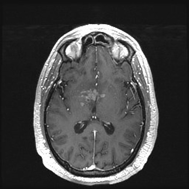
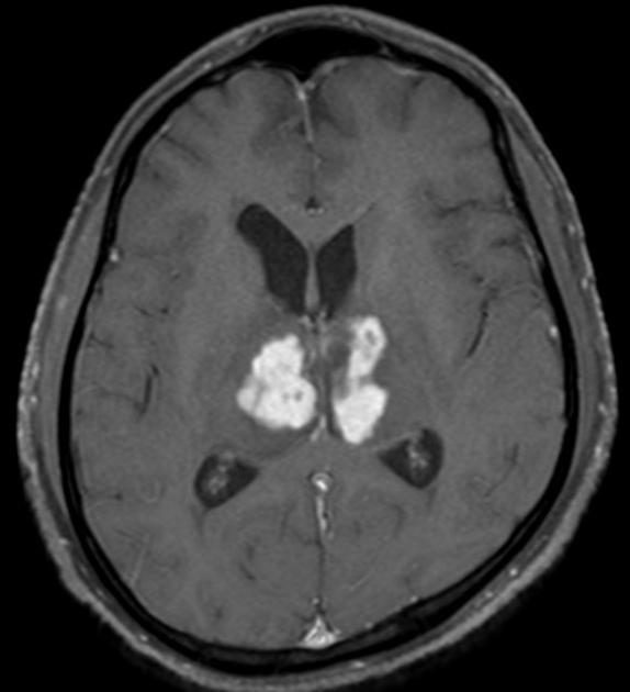
Cerebral amyloidomas are characterised by nodular solid masses with vivid contrast enhancement, typically centred within the white matter, often abutting the lateral ventricles 1. They are often surrounded by a mantle of vasogenic oedema 1. They have most commonly been reported as solitary lesions centred in the white matter of the cerebral hemispheres, although cortical and even posterior fossa lesions have been described 1.
These masses typically grow only slowly if at all, and generally appear to exert less mass effect than one would expect from a similar sized tumour 1.
CT
Amyloidomas generally are somewhat hyperattenuating, although hypoattenuating examples have been reported 1,2. Following administration of contrast, these masses usually enhance 2.
MRI
Signal characteristics of the mass are variable 1,2.
T1: variable signal intensity, ranging from hypointense to hyperintense
T2: variable signal intensity, ranging from hypointense to hyperintense
-
T1 C+
vivid contrast enhancement
peripheral radial enhancement is sometimes seen, thought to correlated with vessel wall cerebral amyloid-β deposition 1
T2*/SWI: microhaemorrhages may be seen
Treatment and prognosis
Cerebral amyloidomas are benign and surgical resection, which is the treatment of choice for symptomatic individuals, is generally curative, although recurrences have been documented 1. Even in asymptomatic individuals, resection or biopsy are usually undertaken as it is difficult to confidently differentiate these lesions from primary or secondary tumours 2. No specific medical therapy has been identified as useful 1.
Differential diagnosis
The differential diagnosis of cerebral amyloidomas is largely that of tumours of the central nervous system, including:


 Unable to process the form. Check for errors and try again.