Cerebral fat embolism
Updates to Article Attributes
Cerebral fat embolism is one manifestation of fat embolism syndrome, but can also rarely occur in isolation.
Epidemiology
Cerebral fat embolism typically occurs in patients with bony fractures (usually long bones of the lower limb) or following orthopaedic or cardiac surgery 19. FatIn particular, fat embolism syndrome has an incidence of 1-3% following long bone fractures and 33% in patients with bilateral long bone fractures 18.
Rarely it has been described as part of a sickle cell crisis with with bone marrow fat necrosis and subsequent embolism 4.
Clinical presentation
Cerebral manifestations of fat embolism syndrome can be differ depending on whether the fat embolism is due to microembolism or macroembolism 19.
-
microembolism
often occurs with fat embolism syndrome
highly variable and non-specific
:clinical presentationthe
symptomsclinical spectrum includes headache, lethargy, irritability, delirium, stupor, convulsions, or coma. Most cases can occur as subclinical events. Concurrentconcurrent pulmonary or cutaneous features may aid in diagnosis
.
-
macroembolism
often occurs without fat embolism syndrome and is a very rare entity
clinical presentation in-keeping with acute ischaemic stroke due to a large vessel occlusion
Pathology
Fat emboli usually reach the brain through either right-to-left cardiac shunt or through intact pulmonary circulation in those without a shunt 3.
Radiographic features
Radiographic features vary depending on whether cerebral fat embolism is due to microembolism or macroembolism.
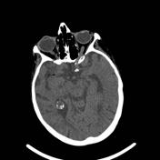
CT
-
ThemicroembolismCT of the brain is normal in most cases 8
. Therethere may be evidence of diffuse oedema with scattered low-attenuating areas and haemorrhage in some situations
.
-
macroembolism
hypodense vessel sign may be present 19
other features in-keeping with ischaemic stroke will eventually develop 19
MRI
TheWith cerebral fat macroembolism, MRI changes are in-keeping with ischaemic stroke from other causes of large vessel occlusion19. However, cerebral fat microembolism has distinct radiographic features on MRI. In cerebral fat microembolism, the distribution of changes in the brain is bilaterally symmetric and predominantly in the subcortical and deep white matter, including subcortical U-fibres, corpus callosum, and internal capsule. SWI and DWI are the most sensitive sequences 17. The distribution and pattern are variable and depends on how extensive embolisation is, but often has an external watershed distribution (similar to other microembolisms).
-
DWI:
early (most common at 1-4 days): scattered punctate foci of cytotoxic oedema (starfield pattern)17
later (most common at 5-14 days): confluent areas of cytotoxic oedema in the white matter 17
SWI: profuse microhaemorrhages in the white matter (walnut kernel pattern)12,13,16,17
T2/FLAIR:
maymay show small areas of high signal intensity indicating vasogenic oedemaT1: corresponding focal regions may show low T1 signal 9
T1 C+:some of the areas of vasogenic may enhance 17
Classification of MRI features
These patterns have been divided into three main types based on chronicity, although the classification system has not been widely adopted in clinical practice 17.
acute (type 1):
scatteredscattered cytotoxic oedema-
subacute (type 2)
type 2A: confluent cytotoxic oedema in white matter
type 2B: vasogenic oedema lesions that may enhance
type 2C: petechial haemorrhages in white matter,
chronic (type 3):
cerebralcerebral atrophychangechange, persistent gliosis
Differential diagnosis
The imaging differential to consider includes many other causes of multiple small foci of infarction or haemorrhage, although generally, only fat emboli will result in a very large number of tiny lesions on both SWI and DWI. Other diagnoses to consider 6:
disseminated intravascular coagulation due to systemic causes other than fat embolisms, such as infection/sepsis
cardiogenic cerebral emboli or septic cerebral emboli
-<p><strong>Cerebral fat embolism </strong>is one manifestation of <a href="/articles/fat-embolism-syndrome">fat embolism syndrome</a>.</p><h4>Epidemiology</h4><p>Cerebral fat embolism typically occurs in patients with bony fractures (usually long bones of the lower limb). Fat embolism syndrome has an incidence of 1-3% following long bone fractures and 33% in patients with bilateral long bone fractures <sup>18</sup>.</p><p>Rarely it has been described as part of a <a href="/articles/sickle-cell-disease-cerebral-manifestations-1">sickle cell crisis</a> with bone marrow fat necrosis and subsequent embolism <sup>4</sup>.</p><h4>Clinical presentation</h4><p>Cerebral manifestations of fat embolism syndrome can be highly variable and non-specific: the symptoms spectrum includes headache, lethargy, irritability, <a href="/articles/delirium">delirium</a>, stupor, convulsions, or coma. Most cases can occur as subclinical events. Concurrent pulmonary or cutaneous features may aid in diagnosis.</p><h4>Pathology</h4><p>Fat emboli usually reach the brain through either right-to-left cardiac shunt or through intact pulmonary circulation in those without a shunt <sup>3</sup>.</p><h4>Radiographic features</h4><h5>CT </h5><p>The CT of the brain is normal in most cases <sup>8</sup>. There may be evidence of diffuse oedema with scattered low-attenuating areas and haemorrhage in some situations.</p><h5>MRI </h5><p>The distribution of changes in the brain is bilaterally symmetric and predominantly in the subcortical and deep white matter, including subcortical U-fibres, corpus callosum, and internal capsule. SWI and DWI are the most sensitive sequences <sup>17</sup>. The distribution and pattern are variable and depends on how extensive embolisation is, but often has an external <a href="/articles/watershed-cerebral-infarction">watershed distribution</a> (similar to other microembolisms). </p><ul>- +<p><strong>Cerebral fat embolism </strong>is one manifestation of <a href="/articles/fat-embolism-syndrome">fat embolism syndrome</a>, but can also rarely occur in isolation.</p><h4>Epidemiology</h4><p>Cerebral fat embolism typically occurs in patients with bony fractures (usually long bones of the lower limb) or following orthopaedic or cardiac surgery <sup>19</sup>. In particular, <a href="/articles/fat-embolism-syndrome">fat embolism syndrome</a> has an incidence of 1-3% following long bone fractures and 33% in patients with bilateral long bone fractures <sup>18</sup>.</p><p>Rarely it has been described as part of a <a href="/articles/sickle-cell-disease-cerebral-manifestations-1">sickle cell crisis</a> with bone marrow fat necrosis and subsequent embolism <sup>4</sup>.</p><h4>Clinical presentation</h4><p>Cerebral manifestations of fat embolism syndrome differ depending on whether the fat embolism is due to microembolism or macroembolism <sup>19</sup>.</p><ul>
-<strong>DWI: </strong><ul>-<li>early (most common at 1-4 days): scattered punctate foci of cytotoxic oedema (<a href="/articles/starfield-pattern-fat-embolism">starfield pattern</a>) <sup>17</sup>-</li>-<li>later (most common at 5-14 days): confluent areas of cytotoxic oedema in the white matter <sup>17</sup>-</li>- +<p>microembolism</p>
- +<ul>
- +<li><p>often occurs with <a href="/articles/fat-embolism-syndrome">fat embolism syndrome</a></p></li>
- +<li><p>highly variable and non-specific clinical presentation</p></li>
- +<li><p>the clinical spectrum includes headache, lethargy, irritability, <a href="/articles/delirium">delirium</a>, stupor, convulsions, or coma</p></li>
- +<li><p>concurrent pulmonary or cutaneous features may aid in diagnosis</p></li>
-<strong>SWI: </strong>profuse microhaemorrhages in the white matter (<a href="/articles/walnut-kernel-microbleed-pattern">walnut kernel pattern</a>) <sup>12,13,16,17</sup>- +<p>macroembolism</p>
- +<ul>
- +<li><p>often occurs without <a href="/articles/fat-embolism-syndrome">fat embolism syndrome</a> and is a very rare entity</p></li>
- +<li><p>clinical presentation in-keeping with acute <a href="/articles/ischaemic-stroke" title="Ischaemic stroke">ischaemic stroke</a> due to a <a href="/articles/large-vessel-occlusion" title="Large vessel occlusion">large vessel occlusion</a></p></li>
- +</ul>
- +</ul><h4>Pathology</h4><p>Fat emboli usually reach the brain through either right-to-left cardiac shunt or through intact pulmonary circulation in those without a shunt <sup>3</sup>.</p><h4>Radiographic features</h4><p>Radiographic features vary depending on whether cerebral fat embolism is due to microembolism or macroembolism.</p><h5>CT </h5><ul>
-<strong>T2/FLAIR:</strong> may show small areas of high signal intensity indicating vasogenic oedema</li>- +<p>microembolism</p>
- +<ul>
- +<li><p>CT of the brain is normal in most cases <sup>8</sup></p></li>
- +<li><p>there may be evidence of diffuse oedema with scattered low-attenuating areas and haemorrhage in some situations</p></li>
- +</ul>
- +</li>
-<strong>T1:</strong> corresponding focal regions may show low T1 signal <sup>9</sup>- +<p>macroembolism</p>
- +<ul>
- +<li><p><a href="/articles/hypodense-vessel-sign" title="Hypodense vessel sign">hypodense vessel sign</a> may be present <sup>19</sup></p></li>
- +<li><p>other features in-keeping with <a href="/articles/ischaemic-stroke" title="Ischaemic stroke">ischaemic stroke</a> will eventually develop <sup>19</sup></p></li>
- +</ul>
- +</ul><h5>MRI </h5><p>With cerebral fat macroembolism, MRI changes are in-keeping with <a href="/articles/ischaemic-stroke" title="Ischaemic stroke">ischaemic stroke</a> from other causes of <a href="/articles/large-vessel-occlusion" title="Large vessel occlusion">large vessel occlusion</a> <sup>19</sup>. However, cerebral fat microembolism has distinct radiographic features on MRI. In cerebral fat microembolism, the distribution of changes in the brain is bilaterally symmetric and predominantly in the subcortical and deep white matter, including subcortical U-fibres, corpus callosum, and internal capsule. SWI and DWI are the most sensitive sequences <sup>17</sup>. The distribution and pattern are variable and depends on how extensive embolisation is, but often has an external <a href="/articles/watershed-cerebral-infarction">watershed distribution</a> (similar to other microembolisms). </p><ul>
-<strong>T1 C+: </strong>some of the areas of vasogenic may enhance <sup>17</sup>- +<p><strong>DWI:</strong></p>
- +<ul>
- +<li><p>early (most common at 1-4 days): scattered punctate foci of cytotoxic oedema (<a href="/articles/starfield-pattern-fat-embolism">starfield pattern</a>) <sup>17</sup></p></li>
- +<li><p>later (most common at 5-14 days): confluent areas of cytotoxic oedema in the white matter <sup>17</sup></p></li>
- +</ul>
-</ul><h6>Classification of MRI features</h6><p>These patterns have been divided into three main types based on chronicity, although the classification system has not been widely adopted in clinical practice <sup>17</sup>. </p><ul>-<li>acute (type 1): scattered cytotoxic oedema</li>-<li>subacute (type 2)<ul>-<li>type 2A: confluent cytotoxic oedema in white matter </li>-<li>type 2B: vasogenic oedema lesions that may enhance </li>-<li>type 2C: petechial haemorrhages in white matter,</li>- +<li><p><strong>SWI: </strong>profuse microhaemorrhages in the white matter (<a href="/articles/walnut-kernel-microbleed-pattern">walnut kernel pattern</a>) <sup>12,13,16,17</sup></p></li>
- +<li><p><strong>T2/FLAIR:</strong> may show small areas of high signal intensity indicating vasogenic oedema</p></li>
- +<li><p><strong>T1:</strong> corresponding focal regions may show low T1 signal <sup>9</sup></p></li>
- +<li><p><strong>T1 C+: </strong>some of the areas of vasogenic may enhance <sup>17</sup></p></li>
- +</ul><h6>Classification of MRI features</h6><p>These patterns have been divided into three main types based on chronicity, although the classification system has not been widely adopted in clinical practice <sup>17</sup>. </p><ul>
- +<li><p>acute (type 1): scattered cytotoxic oedema</p></li>
- +<li>
- +<p>subacute (type 2)</p>
- +<ul>
- +<li><p>type 2A: confluent cytotoxic oedema in white matter </p></li>
- +<li><p>type 2B: vasogenic oedema lesions that may enhance </p></li>
- +<li><p>type 2C: petechial haemorrhages in white matter,</p></li>
-<li>chronic (type 3): cerebral atrophy change, persistent gliosis</li>- +<li><p>chronic (type 3): cerebral atrophy change, persistent gliosis</p></li>
-<li>-<a href="/articles/disseminated-intravascular-coagulation">disseminated intravascular coagulation</a> due to systemic causes other than fat embolisms, such as infection/sepsis</li>-<li><a href="/articles/watershed-cerebral-infarction">watershed infarction</a></li>-<li><a href="/articles/diffuse-axonal-injury-grading-1">diffuse axonal injury</a></li>-<li>cardiogenic cerebral emboli or septic cerebral emboli</li>-<li><a href="/articles/central-nervous-system-vasculitis-2">cerebral vasculitis</a></li>-<li>minute <a href="/articles/haemorrhagic-cerebral-metastases">haemorrhagic cerebral metastases</a>-</li>- +<li><p><a href="/articles/disseminated-intravascular-coagulation">disseminated intravascular coagulation</a> due to systemic causes other than fat embolisms, such as infection/sepsis</p></li>
- +<li><p><a href="/articles/watershed-cerebral-infarction">watershed infarction</a></p></li>
- +<li><p><a href="/articles/diffuse-axonal-injury-grading-1">diffuse axonal injury</a></p></li>
- +<li><p>cardiogenic cerebral emboli or septic cerebral emboli</p></li>
- +<li><p><a href="/articles/central-nervous-system-vasculitis-2">cerebral vasculitis</a></p></li>
- +<li><p>minute <a href="/articles/haemorrhagic-cerebral-metastases">haemorrhagic cerebral metastases</a></p></li>
References changed:
- 14. Bergui M, Castagno D, D’Agata F, Cicerale A, Anselmino M, Ferrio FM, Giustetto C, Halimi F, Scaglione M, Gaita F. Selective Vulnerability of Cortical Border Zone to Microembolic Infarct. (2015) Stroke. <a href="https://doi.org/10.1161/STROKEAHA.114.008194">doi:10.1161/STROKEAHA.114.008194</a> <span class="ref_v4"></span>
- 15. Ryu CW, Lee DH, Kim TK, Kim SJ, Kim HS, Lee JH, Choi CG, Suh DC. Cerebral fat embolism: diffusion-weighted magnetic resonance imaging findings:. (2016) Acta Radiologica. <a href="https://doi.org/10.1080/02841850510021481">doi:10.1080/02841850510021481</a> <span class="ref_v4"></span>
- 19. Ooi S, Raviskanthan S, Campbell B, Hutton E, Mitchell P, Cloud G. Cerebral Large Vessel Occlusion Caused by Fat Embolism—A Case Series and Review of the Literature. Front Neurol. 2021;12:746099. <a href="https://doi.org/10.3389/fneur.2021.746099">doi:10.3389/fneur.2021.746099</a> - <a href="https://www.ncbi.nlm.nih.gov/pubmed/34721272">Pubmed</a>
- 14. Mauro Bergui, Davide Castagno, Federico D’Agata, Alessandro Cicerale, Matteo Anselmino, Federica Maria Ferrio, Carla Giustetto, Franck Halimi, Marco Scaglione, Fiorenzo Gaita. Selective Vulnerability of Cortical Border Zone to Microembolic Infarct. (2015) Stroke. <a href="https://doi.org/10.1161/STROKEAHA.114.008194">doi:10.1161/STROKEAHA.114.008194</a> <span class="ref_v4"></span>
- 15. C. W. Ryu, D. H. Lee, T. K. Kim, S. J. Kim, H. S. Kim, J. H. Lee, C. G. Choi, D. C. Suh. Cerebral fat embolism: diffusion-weighted magnetic resonance imaging findings:. (2016) Acta Radiologica. <a href="https://doi.org/10.1080/02841850510021481">doi:10.1080/02841850510021481</a> <span class="ref_v4"></span>
Image 12 CT (non-contrast) ( create )