
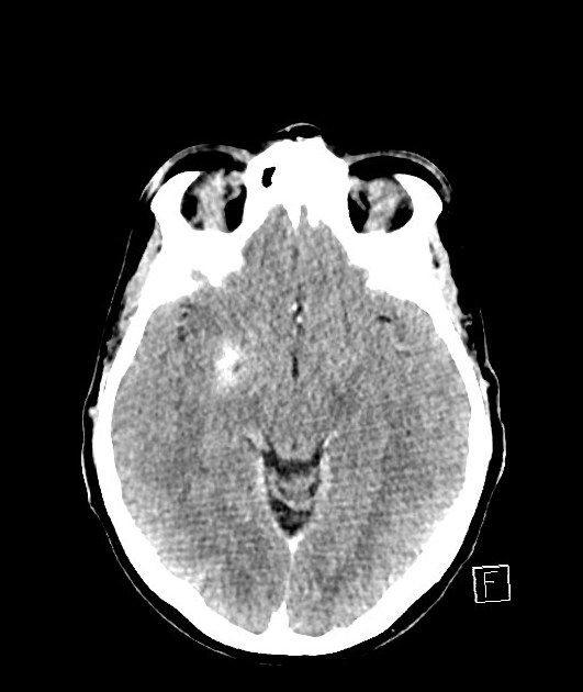
Cerebral intraparenchymal hyperattenuations have been increasingly recognised on CT scans following mechanical thrombectomy for the treatment of thromboembolic ischaemic stroke 1-3. They can also be seen following intravenous thrombolysis (without catheter angiography) 4.
The distribution of cerebral intraparenchymal hyperattenuation correlates with the eventual volume of infarction – in other words, the pre-procedural infarct core plus any portions of penumbra which despite treatment will go on to infarct 1.
As such, intraparenchymal hyperattenuations immediately following mechanical thrombectomy or thrombolysis, observed either on conventional multi-detector CT or flat-panel CT, can provide prognostic information as to the eventual volume of infarction 1.
Terminology
The term is deliberately vague to encompass both contrast staining and petechial haemorrhagic change as distinguishing between the two is not always easy. An alternative term sometimes used is postinterventional cerebral hyperdensity 2.
Radiographic features
Distinguishing between contrast staining and petechial haemorrhagic transformation is not easy, particularly when obtained on flat-panel CT.
Contrast staining
Contrast staining typically appears as areas of hyperdensity mostly confined to grey matter (cortex and deep grey matter) and represent areas of blood-brain barrier breakdown secondary to ischaemia with microvascular extravasation of contrast into the extracellular space 1. Typically such staining clears within the first 19-24 hours after the procedure 2.
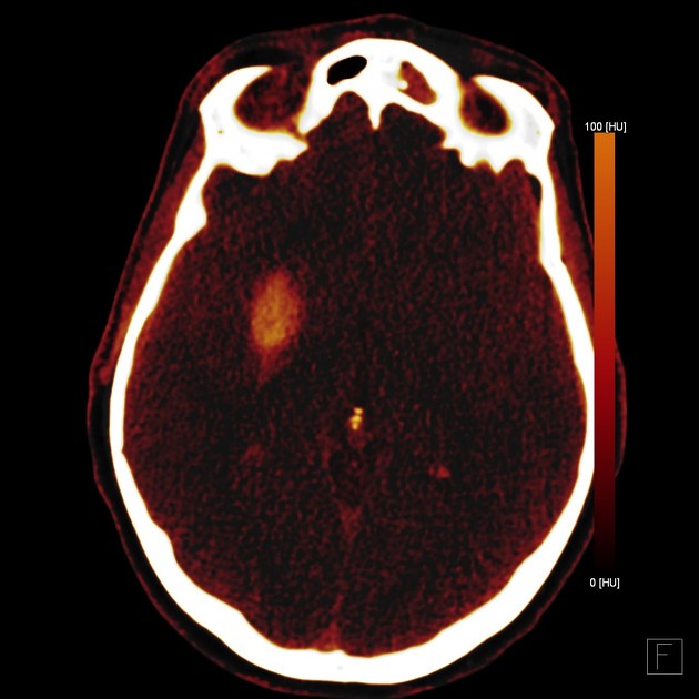
Dual-energy CT is also able to distinguish hyperdensity due to contrast versus hyperdensity due to haemorrhage 3.
Petechial haemorrhagic transformation
Petechial haemorrhagic transformation of an ischaemic infarct (as opposed to macroscopic solid cerebral haemorrhages also sometimes encountered) can have a very similar appearance. Follow-up CT performed at least 19-24 hours following intervention is the most specific way to differentiate, with persistent hyperdensity consistent with haemorrhage whereas contrast staining will reduce in density over time 2.


 Unable to process the form. Check for errors and try again.