
The differential for peripheral or ring enhancing cerebral lesions includes:
tumefactive demyelinating lesion (incomplete ring)
postoperative change
lymphoma - in an immunocompromised patient
necrotising leukoencephalopathy after methotrexate 4,5
A helpful mnemonic is MAGIC DR.
On this page:
Radiographic features
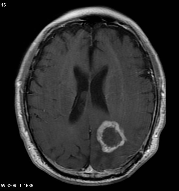
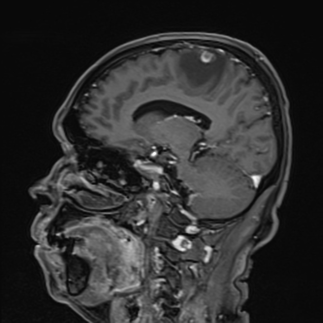
No single feature is pathognomonic, although a cystic lesion that markedly restricts centrally (the fluid component) on DWI should be considered an abscess until proven otherwise.
Many features of the lesion, as well as clinical presentation and patient demographics, need to be taken together to help narrow the differential. Helpful rules of thumb include:
-
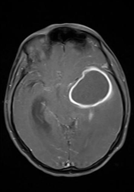
enhancing wall characteristics
thick and nodular favours neoplasm
thin and regular favours abscess
incomplete ring often opened toward the cortex favours demyelination
intermediate to low T2 signal capsule favours abscess
restricted diffusion of enhancing wall favours GBM or demyelination
-
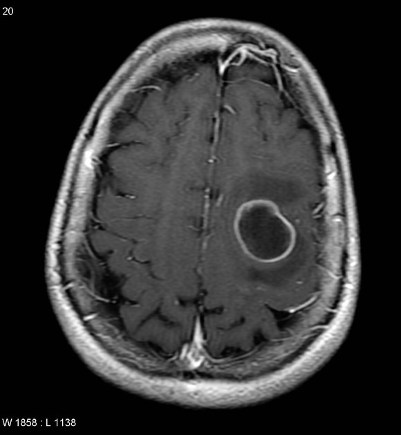
surrounding oedema
extensive oedema relative to lesion size favours abscess
increased perfusion favours neoplasm (metastases or primary cerebral malignancy)
-
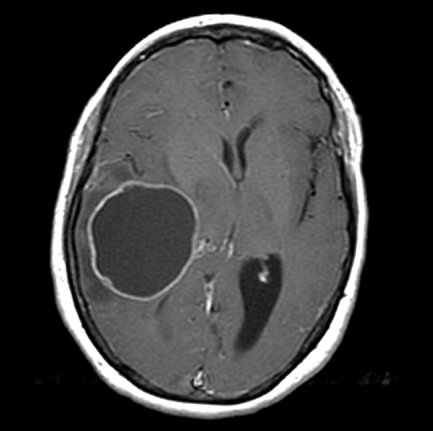
central fluid content
restricted diffusion favours abscess
an absence of diffusion restriction favour a tumour with a central necrotic component (classically metastases)
-
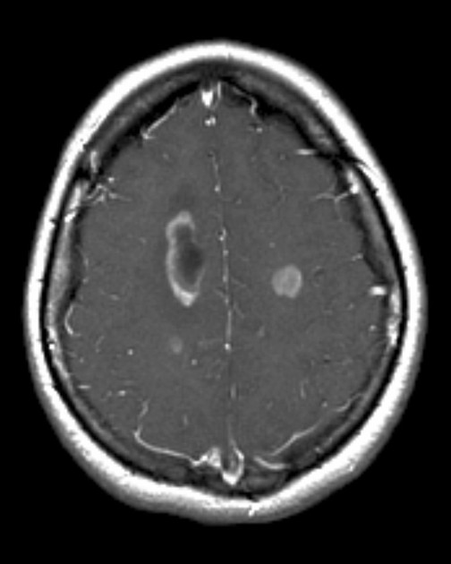
number of lesions
similar sized rounded lesions at grey-white matter junction favours metastases or abscesses
irregular mass with adjacent secondary lesions embedded in the same region of 'oedema' favours GBM
small (<1-2 cm) lesions with thin walls, especially if other calcific foci are present, suggest neurocysticercosis.


 Unable to process the form. Check for errors and try again.