
Ciliary body (eye)
Citation, DOI, disclosures and article data
At the time the article was created Arjun Raju had no financial relationships to ineligible companies to disclose.
View Arjun Raju's current disclosuresAt the time the article was last revised Craig Hacking had the following disclosures:
- Philips Australia, Paid speaker at Philips Spectral CT events (ongoing)
- Taylor and Francis Publishing, Paid author of Imaging for Students 5th edition (ongoing)
These were assessed during peer review and were determined to not be relevant to the changes that were made.
View Craig Hacking's current disclosures- ciliary bodies
The ciliary body is the continuation of the uveal layer of the eye and functions in the production of aqueous humour and the process of lens accommodation.
On this page:
Summary
location: between the vitreous body and posterior chamber of the globe
function: aqueous humour production and accommodation of the lens
arterial supply: anterior ciliary arteries and long posterior ciliary arteries
venous drainage: vorticose veins
innervation: short ciliary nerves
relations: vitreous cavity posteriorly, posterior chamber anteriorly, scleral layer externally, retinal layer internally
Gross anatomy
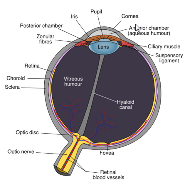
The ciliary body is a continuation of the uveal tract, with the choroid lying posteriorly and iris anteriorly. The uveal tract lies between an outer scleral layer and inner retina layer of the eye 1. It forms a flat ring and contains the ciliary process, ciliary muscles and ciliary vessels.
The ciliary processes are finger like protrusions of the ciliary body and attach to the lens via the zonula fibres (suspensory ligaments), allowing for the process of accommodation. The ciliary processes contain specialised vascular epithelium that secrete aqueous humour into the globe of the eye 1.
The ciliary muscles consist of three differently orientated smooth muscle fibres. The outermost longitudinal fibres attach the ciliary body to the scleral spur and function to open the trabecular network and Schlemm’s canal 2. The innermost circular fibres relax the zonular fibres when contracted, allowing for an increase in lens axial diameter and convexity. Radial or oblique fibres lie in between and connect the two layers 2.
The ciliary body is supplied by the anterior ciliary arteries and the long posterior ciliary arteries which anastomose and form a vascular ring around the root of the iris. It is drained by the vorticose veins and then into the superior and inferior orbital veins.
The ciliary body receives parasympathetic innervation from the Edinger-Westphal nucleus in the midbrain and travels via the oculomotor nerve to the ciliary ganglion. This ganglion gives off short ciliary nerves which innervates the ciliary body.
Histology
The ciliary body contains two layers of cuboidal epithelium. The deep layer is heavily pigmented due to high vascularity and high level of melanin. The surface layer is non-pigmented, non-photosensitive extension of the receptor layer of the retina and is responsible for aqueous humour production 3.
Related pathology
open-angle glaucoma
angle-closure glaucoma
panophthalmitis
ciliary body melanoma
References
- 1. Chummy S. Sinnatamby. Last's Anatomy. (2011) ISBN: 9780702033957 - Google Books
- 2. Borges- Giampani A & Giampani J. Anatomy of Ciliary Body, Ciliary Processes, Anterior Chamber Angle and Collector Vessels. Glaucoma - Basic and Clinical Aspects. 2013. doi:10.5772/52780
- 3. Barbara Young, Geraldine O'Dowd, Phillip Woodford. Wheater's Functional Histology. (2013) ISBN: 9780702047473 - Google Books
Incoming Links
Related articles: Anatomy: Head and neck
- skeleton of the head and neck[+][+]
-
cranial vault
- scalp (mnemonic)
- fontanelle
-
sutures
- calvarial
- facial
- frontozygomatic suture
- frontomaxillary suture
- frontolacrimal suture
- frontonasal suture
- temporozygomatic suture
- zygomaticomaxillary suture
- parietotemporal suture (parietomastoid suture)
- occipitotemporal suture (occipitomastoid suture)
- sphenofrontal suture
- sphenozygomatic suture
- spheno-occipital suture (not a true suture)
- lacrimomaxillary suture
- nasomaxillary suture
- internasal suture
- basal/internal
- skull landmarks
- frontal bone
- temporal bone
- parietal bone
- occipital bone
- skull base (foramina)
-
facial bones
- midline single bones
- paired bilateral bones
- cervical spine
- hyoid bone
- laryngeal cartilages
-
cranial vault
- muscles of the head and neck[+][+]
- muscles of the tongue (mnemonic)
- muscles of mastication
-
facial muscles
- epicranius muscle
- circumorbital and palpebral muscles
- nasal muscles
-
buccolabial muscles
- elevators, retractors and evertors of the upper lip
- levator labii superioris alaeque nasalis muscle
- levator labii superioris muscle
- zygomaticus major muscle
- zygomaticus minor muscle
- levator anguli oris muscle
- malaris muscle
- risorius muscle
- depressors, retractors and evertors of the lower lip
- depressor labii inferioris muscle
- depressor anguli oris muscle
- mentalis muscle
- compound sphincter
-
orbicularis oris muscle
- incisivus labii superioris muscle
- incisivus labii inferioris muscle
-
orbicularis oris muscle
- muscle of mastication
- modiolus
- elevators, retractors and evertors of the upper lip
- muscles of the middle ear
- orbital muscles
- muscles of the soft palate
- pharyngeal muscles
- suprahyoid muscles
- infrahyoid muscles
- intrinsic muscles of the larynx
- muscles of the neck
- platysma muscle
- longus colli muscle
- longus capitis muscle
- scalenus anterior muscle
- scalenus medius muscle
- scalenus posterior muscle
- scalenus pleuralis muscle
- sternocleidomastoid muscle
-
suboccipital muscles
- rectus capitis posterior major muscle
- rectus capitis posterior minor muscle
- obliquus capitis superior muscle
- obliquus capitis inferior muscle
- accessory muscles of the neck
- deep cervical fascia[+][+]
-
deep spaces of the neck[+][+]
- anterior cervical space
- buccal space
- carotid space
- danger space
- deep cervical fascia
- infratemporal fossa
- masticator space
- parapharyngeal space
- stylomandibular tunnel
- parotid space
- pharyngeal (superficial) mucosal space
- perivertebral space
- posterior cervical space
- pterygopalatine fossa
- retropharyngeal space
- suprasternal space (of Burns)
- visceral space
- surgical triangles of the neck[+][+]
- orbit
- ear[+][+]
- paranasal sinuses[+][+]
- upper respiratory tract[+][+]
- viscera of the neck[+][+]
- blood supply of the head and neck[+][+]
-
arterial supply
-
common carotid artery
- carotid body
- carotid bifurcation
- subclavian artery
- variants
-
common carotid artery
- venous drainage
-
arterial supply
- innervation of the head and neck[+][+]
-
cranial nerves
- olfactory nerve (CN I)
- optic nerve (CN II)
- oculomotor nerve (CN III)
- trochlear nerve (CN IV)
-
trigeminal nerve (CN V) (mnemonic)
- trigeminal ganglion
- ophthalmic division
- maxillary division
- mandibular division
- abducens nerve (CN VI)
- facial nerve (CN VII)
-
vestibulocochlear nerve (CN VIII)
- vestibular ganglion (Scarpa's ganglion)
- glossopharyngeal nerve (CN IX)
- vagus nerve (CN X)
- (spinal) accessory nerve (CN XI)
- hypoglossal nerve (CN XII)
- parasympathetic ganglia of the head and neck
- cervical sympathetic ganglia
- greater occipital nerve
- third occipital nerve
-
cervical plexus
- muscular branches
- longus capitis
- longus colli
- scalenes
- geniohyoid
- thyrohyoid
-
ansa cervicalis
- omohyoid (superior and inferior bellies separately)
- sternothyroid
- sternohyoid
- phrenic nerve
- contribution to the accessory nerve (CN XI)
- cutaneous branches
- muscular branches
- brachial plexus
- pharyngeal plexus
-
cranial nerves
- lymphatic drainage of the head and neck[+][+]
- embryological development of the head and neck[+][+]


 Unable to process the form. Check for errors and try again.