
Coccydynia refers to pain in and among the area of the coccyx. It is characterised by coccygeal pain which is typically provocated by pressure. It may remain unclear in origin owing to the unpredictability of the source of pain 1. Most of patient presented wth coccydynia had prior history of fall, particularly in sitting position12.
On this page:
Epidemiology
No accurate data about the frequency of coccydynia is reported, but one study found that coccydynia accounted for ~3% of all low back pain hospital presentations 8. It is ~5x more common in females than males ref.
Risk factors
obesity 10
female sex 10
osteoporosis and other degenerative bone diseases ref
osteomyelitis ref
contact sports ref
elderly with risk of fall
Associations
obesity ref
Clinical presentation
As the name suggests, pain is considered to be the primary symptom of coccydynia. However, pain is extremely varied and may be related to sitting, bowel movements, radiculopathic or generalised coccygeal pain. The coccyx may be tender on examination.
Pathology
Aetiology
Coccydynia can be classified into ref:
idiopathic (most common) ref
-
secondary coccygeal pain
-
abnormal coccygeal mobility
hypermobility 10
rigid coccyx with spicule at the tip 10
intersegmental coccygeal subluxation 10
-
trauma
post falls or trauma to gluteal region 10
-
childbirth, particularly instrumented or difficult deliveries 13
both subluxation and uncommonly fractures can occur 13
partial dislocation of the sacrococcygeal synchondrosis
intersegmental degeneration 10
tumour, e.g. chordomas, chondrosarcoma (rare) 10
infection, e.g. tuberculosis (rare) 10
-
Radiographic features
Coccydynia is essentially a clinical diagnosis, but imaging modalities are helpful in assessment and possible identification of the aetiology.
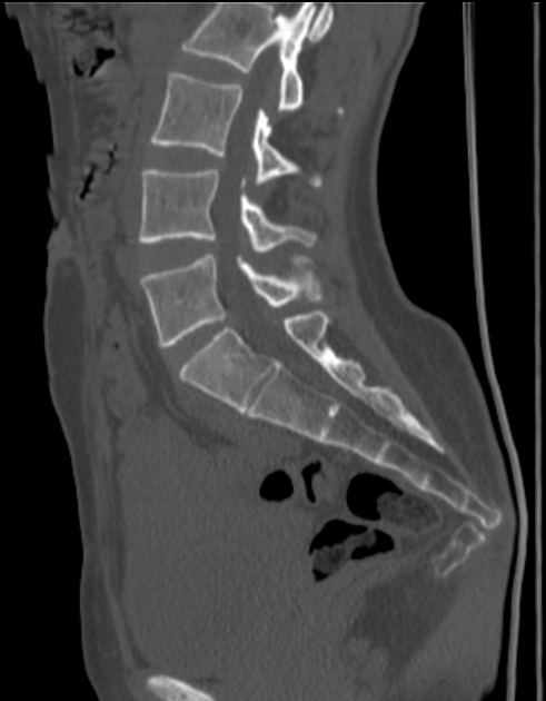
Plain radiograph
Considered the first line of imaging of painful coccyx 10. Two types of radiographs:
-
standard radiograph
-
coccyx scoliotic deformity (AP and lateral)
number and morphology of coccygeal segments including degenerative change
useful radiological assessment to evaluate anterior angulation of the coccyx and its deformity
-
-
dynamic sitting and standing radiographs 10
superior to standard radiograph because it can detect measurement of the sagittal rotation of the pelvis and the coccygeal angle of incidence ref
-
allows for assessment of coccygeal mobility by measuring the intercoccygeal angle with the generally accepted values being (although this is not validated/standardised) 10
dynamic posterior intersegmental coccygeal subluxation 10
<5°: immobile/rigid 10
5° to 25°: normal 10
>25°: hypermobility 7,10
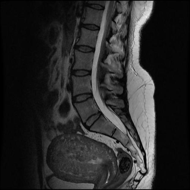
MRI
Described features include 11:
rigid coccyx with a spicule or spur at its tip
bursa along the dorsal surface of the coccyx
presence of fluid collection within the sacrococcygeal synchondrosis
large draining vein on the ventral coccyx
any inflammation or soft tissue abnormalities around the coccyx
Treatment and prognosis
Conservative treatment includes rest, coccygeal cushion, physiotherapy and massage. In traumatic coccydynia the joint may heal spontaneously over weeks or months
Interventions include:
injections of local anaesthetic and steroid
radiofrequency ablation of coccygeal discs and ganglion impar
-
removal of the coccyx by surgery either partial or total (coccygectomy) in refractory cases
limited coccygectomy may to resect a mobile segment 5
History and etymology
The word coccyx originates from the Greek word "cuckoo" (kokkyx), on the basis of resemblance to the structure a cuckoo's beak. The term was used in practice for the first time by Simpson in 1859, although was described as early as 1600s. It is also called the "tailbone" because it is located anatomically at the end of the vertebral column below the sacrum.
Differential diagnosis
Causes of pain in the coccygeal region include:
pilonidal sinus/abscess
sciatica
infection including shingles of the buttocks


 Unable to process the form. Check for errors and try again.