
Colonic pseudo-obstruction, also known as Ogilvie syndrome, is a potentially fatal condition leading to an acute colonic distention without an underlying mechanical obstruction. It is defined as an acute pseudo-obstruction and dilatation of the colon in the absence of any mechanical obstruction.
On this page:
Epidemiology
Numerous causes have been identified, and the demographics of affected patients generally reflect these, with elderly unwell patients being most frequently affected 4. Usually seen in people over 60 years of age and there is a male predilection 5.
Clinical presentation
Patients usually present with constipation, nausea, vomiting and abdominal distension. Colonic pseudo-obstruction can present with a sudden painless enlargement of the proximal colon accompanied by distension. Bowel sounds are normal or high-pitched, but should not be absent.
Despite the absence of mechanical obstruction, patients can nonetheless go on to bowel necrosis and perforation (especially if dilatation is severe) which in turn can go on to become generalized peritonitis 3.
Pathology
Colonic pseudo-obstruction is related to decreased parasympathetic activity.
Risk factors include ref:
trauma
burns
recent surgery
-
medications
opioids
phenothiazines
clozapine
respiratory failure
electrolyte disturbances
uremia
neurological disease (e.g. Parkinson disease, paraneoplastic neuropathy) 8,9
postpartum (particularly post-Cesarean section delivery) 10
Radiographic features
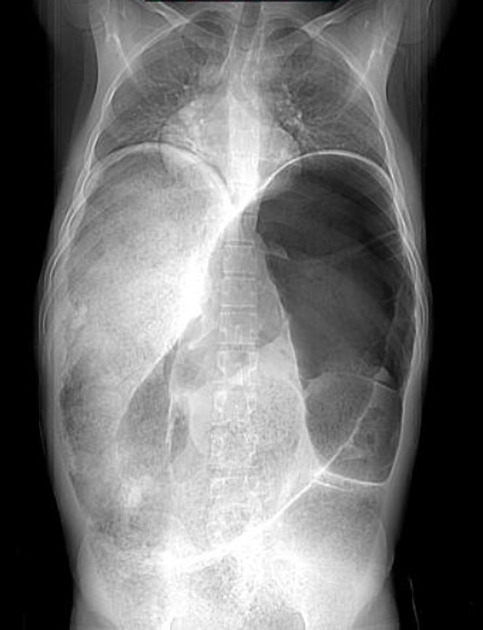
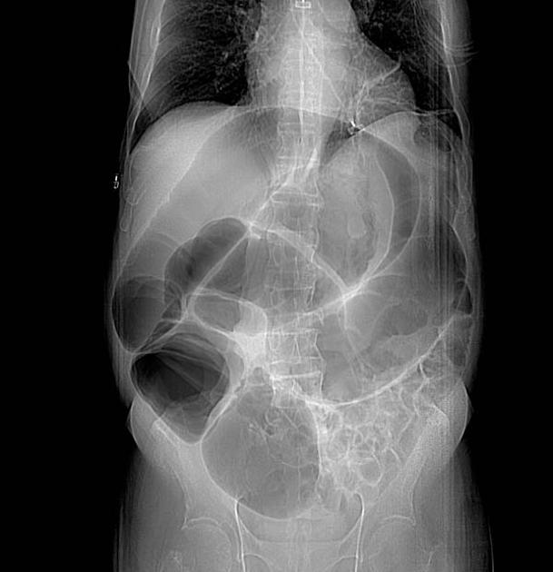
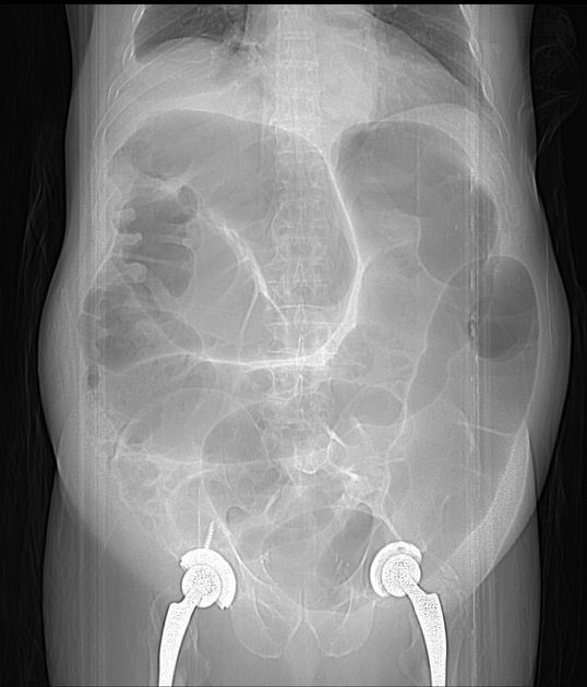
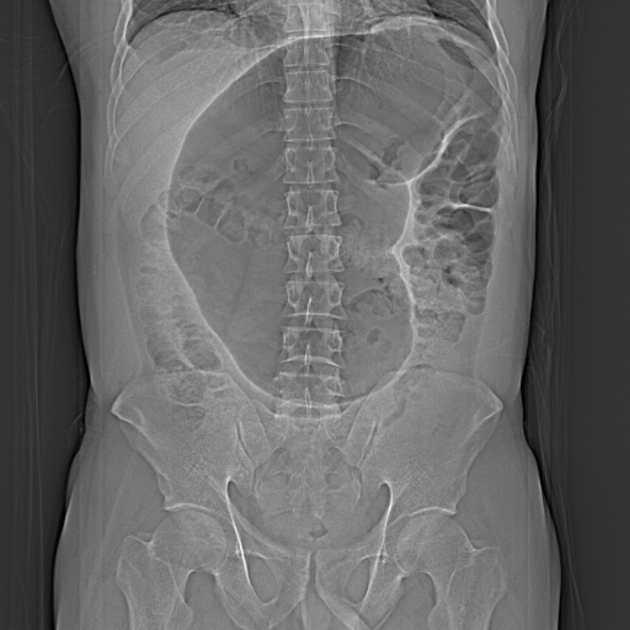
Plain radiograph
Findings can be identical to a mechanical large bowel obstruction 1.
Fluoroscopy
A single contrast/water-soluble enema demonstrates the absence of any mechanical obstruction.
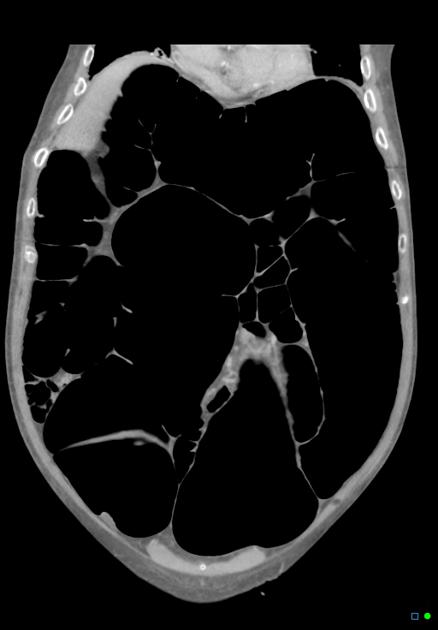
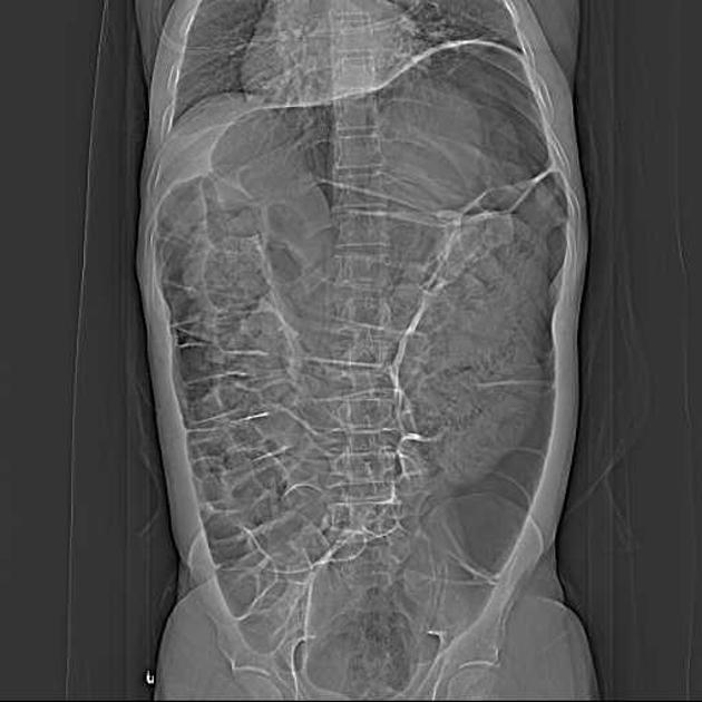
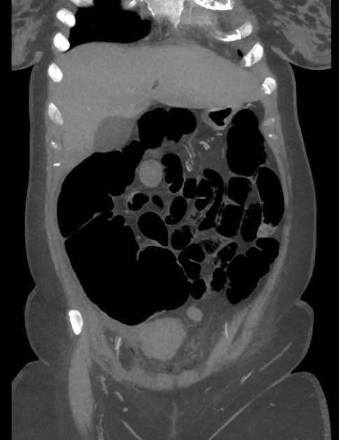
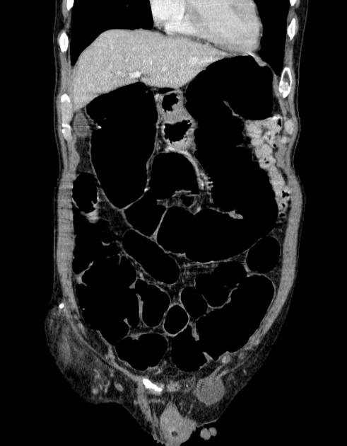
CT
The hallmark of colonic pseudo-obstruction is the presence of dilatation of the large bowel (often marked) without evidence of an abrupt transition point or mechanically obstructing lesion.
It is important to note, however, that a gradual transition point is frequently present, usually at or near the splenic flexure 3.
Treatment and prognosis
Treatment involves correction of the underlying disorder and correction of any biochemical abnormalities. Medical treatment options include anticholinesterases like neostigmine and antibiotics like erythromycin 5,11.
Decompression with a rectal tube (endoscopic decompression) or careful colonoscopy may be effective 7. Cecal distension over 11 cm is predictive of medical/endoscopic management failure 11. In severe cases, surgical or fluoroscopy-assisted cecostomy is recommended, or even occasionally a percutaneous endoscopic colostomy (PEC) 5,11.
Complications
Cecal perforation (more than one quarter perforate when cecal diameter is over 12 cm) 5
Organ failure
History and etymology
It was initially described by the British general surgeon Sir William Heneage Ogilvie (1887-1971) in 1948 1,6.
Differential diagnosis
General imaging differential considerations include:
-
no transition point
often history has a cause for the ileus, e.g. surgery
small bowel is also often dilated
-
mechanical large bowel obstruction
abrupt transition point often with an identifiable obstructing lesion
-
toxic megacolon secondary to Clostridioides difficile colitis
C. difficile infection is usually preceded by antibiotic use or chemotherapy and is therefore usually encountered in unwell, hospitalized patients with significant co-morbidity
bowel wall thickening usually a prominent feature
-
usually bowel wall is thickened, but can be thinned and dilated
absent/poor wall enhancement
usually involves vascular territories
-
sigmoid volvulus and cecal volvulus
transition point evident
whirlpool sign of the twisted mesentery


 Unable to process the form. Check for errors and try again.