
Condylar canal
Citation, DOI, disclosures and article data
Citation:
Mudgal P, Sharma R, Catalá E, et al. Condylar canal. Reference article, Radiopaedia.org (Accessed on 17 Mar 2025) https://doi.org/10.53347/rID-28229
rID:
28229
Article created:
16 Mar 2014,
Prashant Mudgal
Disclosures:
At the time the article was created Prashant Mudgal had no recorded disclosures.
View Prashant Mudgal's current disclosures
Last revised:
Disclosures:
At the time the article was last revised Rohit Sharma had no financial relationships to ineligible companies to disclose.
View Rohit Sharma's current disclosures
Revisions:
14 times, by
10 contributors -
see full revision history and disclosures
Systems:
Sections:
Synonyms:
- Canalis condylaris
- Posterior condylar canal
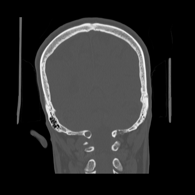
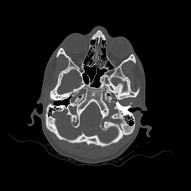
The condylar canal, or canalis condylaris, is a skull base canal in the posterior cranial fossa, located in the condylar fossa. It is the largest of the emissary foramina of the skull 1.
Summary
- location: in the condylar fossa of the posterior cranial fossa, posterior to the occipital condyles
-
contents
- emissary veins, connecting the sigmoid sinus to the occipital vein
- meningeal branch of the occipital artery
Variant anatomy
The condylar canal has a variable presence and is seen only in ~55% (range 50-60%) of cases. It is more commonly bilateral.
Quiz questions
References
- 1. Ginsberg LE. The posterior condylar canal. AJNR Am J Neuroradiol. 1994;15 (5): 969-72. AJNR Am J Neuroradiol (abstract) - Pubmed citation
- 2. Schulte E. Thieme Atlas of Anatomy. Thieme Georg Verlag. (2007) ISBN:3131421010. Read it at Google Books - Find it at Amazon
Incoming Links
Cases:
Multiple choice questions:
Related articles: Anatomy: Head and neck
- skeleton of the head and neck
-
cranial vault [+][+]
- scalp (mnemonic)
- fontanelle
-
sutures
- calvarial
- facial
- frontozygomatic suture
- frontomaxillary suture
- frontolacrimal suture
- frontonasal suture
- temporozygomatic suture
- zygomaticomaxillary suture
- parietotemporal suture (parietomastoid suture)
- occipitotemporal suture (occipitomastoid suture)
- sphenofrontal suture
- sphenozygomatic suture
- spheno-occipital suture (not a true suture)
- lacrimomaxillary suture
- nasomaxillary suture
- internasal suture
- basal/internal
- skull landmarks
- frontal bone
- temporal bone
- parietal bone
- occipital bone
- skull base (foramina)
-
facial bones[+][+]
- midline single bones
- paired bilateral bones
- cervical spine
- hyoid bone
- laryngeal cartilages[+][+]
-
cranial vault [+][+]
- muscles of the head and neck[+][+]
- muscles of the tongue (mnemonic)
- muscles of mastication
-
facial muscles
- epicranius muscle
- circumorbital and palpebral muscles
- nasal muscles
-
buccolabial muscles
- elevators, retractors and evertors of the upper lip
- levator labii superioris alaeque nasalis muscle
- levator labii superioris muscle
- zygomaticus major muscle
- zygomaticus minor muscle
- levator anguli oris muscle
- malaris muscle
- risorius muscle
- depressors, retractors and evertors of the lower lip
- depressor labii inferioris muscle
- depressor anguli oris muscle
- mentalis muscle
- compound sphincter
-
orbicularis oris muscle
- incisivus labii superioris muscle
- incisivus labii inferioris muscle
-
orbicularis oris muscle
- muscle of mastication
- modiolus
- elevators, retractors and evertors of the upper lip
- muscles of the middle ear
- orbital muscles
- muscles of the soft palate
- pharyngeal muscles
- suprahyoid muscles
- infrahyoid muscles
- intrinsic muscles of the larynx
- muscles of the neck
- platysma muscle
- longus colli muscle
- longus capitis muscle
- scalenus anterior muscle
- scalenus medius muscle
- scalenus posterior muscle
- scalenus pleuralis muscle
- sternocleidomastoid muscle
-
suboccipital muscles
- rectus capitis posterior major muscle
- rectus capitis posterior minor muscle
- obliquus capitis superior muscle
- obliquus capitis inferior muscle
- accessory muscles of the neck
- deep cervical fascia[+][+]
-
deep spaces of the neck[+][+]
- anterior cervical space
- buccal space
- carotid space
- danger space
- deep cervical fascia
- infratemporal fossa
- masticator space
- parapharyngeal space
- stylomandibular tunnel
- parotid space
- pharyngeal (superficial) mucosal space
- perivertebral space
- posterior cervical space
- pterygopalatine fossa
- retropharyngeal space
- suprasternal space (of Burns)
- visceral space
- surgical triangles of the neck[+][+]
- orbit[+][+]
- ear[+][+]
- paranasal sinuses[+][+]
- upper respiratory tract[+][+]
- viscera of the neck[+][+]
- blood supply of the head and neck[+][+]
-
arterial supply
-
common carotid artery
- carotid body
- carotid bifurcation
- subclavian artery
- variants
-
common carotid artery
- venous drainage
-
arterial supply
- innervation of the head and neck[+][+]
-
cranial nerves
- olfactory nerve (CN I)
- optic nerve (CN II)
- oculomotor nerve (CN III)
- trochlear nerve (CN IV)
-
trigeminal nerve (CN V) (mnemonic)
- trigeminal ganglion
- ophthalmic division
- maxillary division
- mandibular division
- abducens nerve (CN VI)
- facial nerve (CN VII)
-
vestibulocochlear nerve (CN VIII)
- vestibular ganglion (Scarpa's ganglion)
- glossopharyngeal nerve (CN IX)
- vagus nerve (CN X)
- (spinal) accessory nerve (CN XI)
- hypoglossal nerve (CN XII)
- parasympathetic ganglia of the head and neck
- cervical sympathetic ganglia
- greater occipital nerve
- third occipital nerve
-
cervical plexus
- muscular branches
- longus capitis
- longus colli
- scalenes
- geniohyoid
- thyrohyoid
-
ansa cervicalis
- omohyoid (superior and inferior bellies separately)
- sternothyroid
- sternohyoid
- phrenic nerve
- contribution to the accessory nerve (CN XI)
- cutaneous branches
- muscular branches
- brachial plexus
- pharyngeal plexus
-
cranial nerves
- lymphatic drainage of the head and neck[+][+]
- embryological development of the head and neck[+][+]


 Unable to process the form. Check for errors and try again.