
Congenital aortic stenosis broadly refers to a congenital narrowing of the aortic lumen. Although the term can mean narrowing at any point, it often relates to a narrowing of the aortic valve. As a broad group, there can be some overlap with ascending aortic coarctation depending on the definition used.
On this page:
Epidemiology
Associations
Williams syndrome: with supravalvular type
Pathology
Depending on location it can be classified into three types:
supravalvular stenosis
congenital aortic valve stenosis (commonest)
subvalvular stenosis
Radiographic features
Plain radiograph
Chest radiographs can be normal or may show evidence of cardiomegaly.
Ultrasound
Echocardiography
Echocardiograms may show a high flow jet through the aortic valve or a narrowed segment.
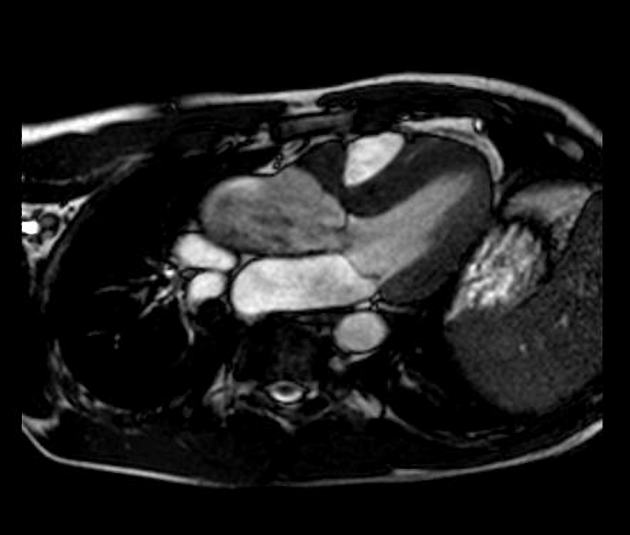
CT/MRI
Apart from showing a narrowed valve annulus and/or narrowing cross-sectional aortic segment, it may also show:
cardiomegaly with left ventricular hypertrophy
post-stenotic dilated segment of the aortic lumen
On MR imaging, velocity encoded phase-contrast cine sequences can assist in assessing the severity of the stenosis by allowing measurement of blood flow velocities and volumes 2.


 Unable to process the form. Check for errors and try again.