
CT enterography (CTE) is a non-invasive technique for diagnosing small bowel disorders.
On this page:
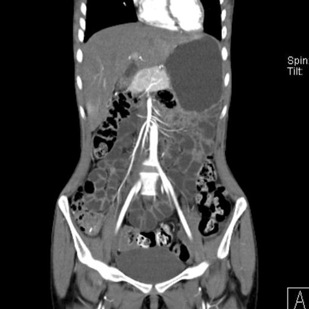
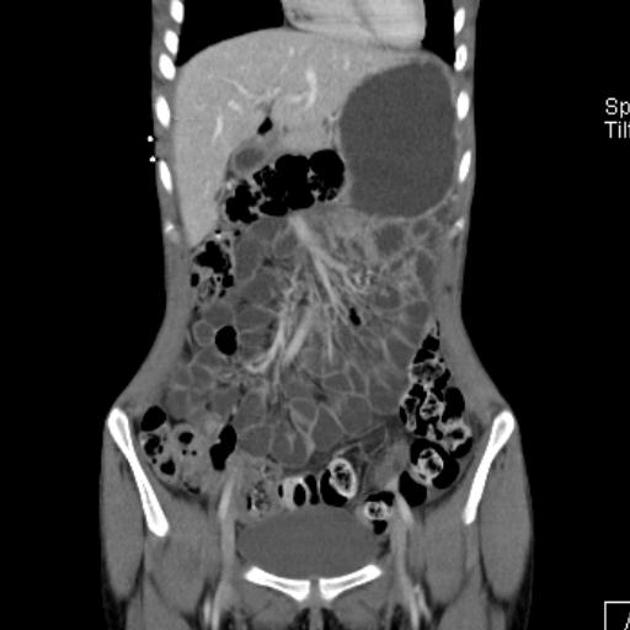
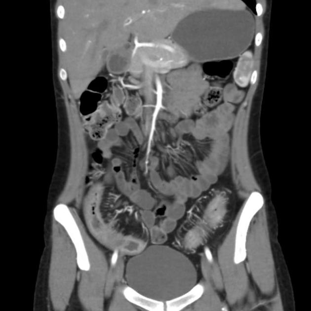
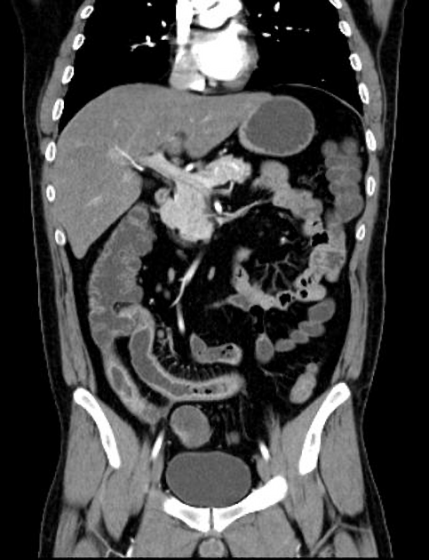
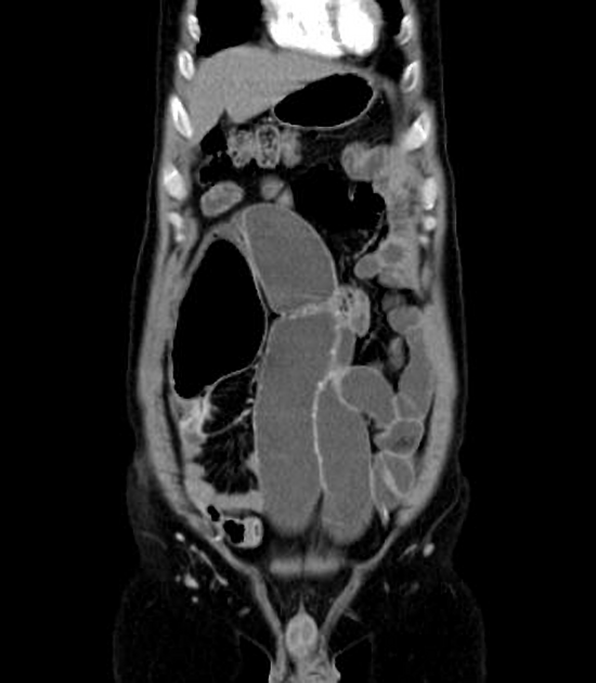
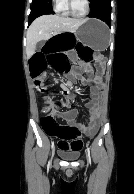
Images:
Indications
Indications for CT enterography include 4,8:
-
diagnosis and complications (primarily)
most common indication
suspected small bowel bleeding, usually performed after a negative endoscopy
suspected small bowel tumour, e.g. neuroendocrine tumour (NET), polyposis syndromes
coeliac disease: assess for complications such as lymphoma
partial small bowel obstructions, e.g. postoperative adhesions, radiation enteritis, scleroderma
chronic diarrhoea and/or abdominal pain
suspected chronic mesenteric ischaemia
Advantages
useful in the assessment of the solid organs and provides a global overview of the abdomen 1
Disadvantages
exposure to ionising radiation
Technique
NB: This article is intended to outline some general principles of protocol design. The specifics will vary depending on CT hardware and software, radiologist's and referrer's preferences, institutional protocols, and patient factors (e.g. allergy and fluid intake restrictions).
Bowel preparation
abstain from all food and drink 4-6 hours before the exam
-
patients drink about 1.5 L of oral contrast over 30-60 minute
adequate luminal distension is necessary as collapsed bowel loops may mimic pathology
-
CT enterography utilises negative or neutral oral contrast 1-3
attenuation similar to that of water - e.g. water, PEG, mannitol, methylcellulose, locust bean gum, and low-density barium sulphate preparations (Volumen, 0.1% W/V)
Fluid distension of the small bowel allows better assessment of mucosal enhancement, mural thickness as well as mesenteric vasculature, this is important especially in the evaluation of Crohn disease 2.
Procedure
CT scanning is ideally performed on a multidetector computed tomography (MDCT) scanner
-
intravenous contrast
Crohn disease, coeliac disease, postoperative adhesions, radiation enteritis, and scleroderma: a single enteric phase where peak mucosal enhancement is achieved is sufficient - either enteric phase (45-50s) or portal venous phase (60-70s)
small bowel tumours: an additional arterial phase can be performed, in particular for the assessment of hypervascular lesions (e.g. NETs)
in cases of suspected GI bleeding, pre-contrast, arterial, portal venous, and delayed phases should be considered
data interpretation with the use of axial and coronal reformatted images for proper evaluation
Findings
inflammatory bowel disease and its complications e.g. Crohn disease or ulcerative colitis
small bowel tumours, including benign tumours (e.g. hamartomatous or hyperplastic polyps) or malignant tumours (e.g. adenocarcinoma, NET, lymphoma, gastrointestinal stromal tumours)
mesenteric ischaemia and gastrointestinal tract bleeding
Coeliac disease 1,2


 Unable to process the form. Check for errors and try again.