
Presentation
Abdominal pain and diarrhea.
Patient Data
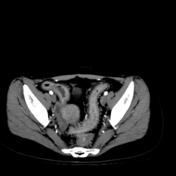


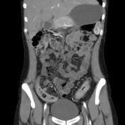
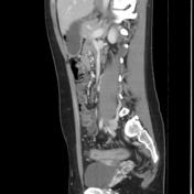

The terminal ileum, the sigmoid and the descending colon show mural thickening and contrast enhancement of the mucosa.
Prominent pericentric and pericolic vasculature highlighted by fibrofatty proliferation of the mesentery.
Vascular dilatation and tortuosity of the vasa recta, giving the comb sign.
Few enlarged pericolic and mesenteric lymph nodes.
Case Discussion
This is an endoscopy and biopsy-proven case of "Crohn's disease"
Crohn's disease, like ulcerative colitis, is an idiopathic inflammatory bowel disease (IBD), characterized by widespread gastrointestinal tract involvement, typically with skip lesions - thereby its synonym regional enteritis - and frequent systemic involvement.
CT examination can be carried out with both intravenous and intraluminal contrast (positive or negative).
bowel wall enhancement
bowel wall thickening (1-2 cm) which is most frequently seen in the terminal ileum (present in up to 83% of patients)
strictures and fistulae
mesenteric / intra-abdominal abscess or phlegmon formation
abscesses are eventually seen in 15-20% of patients
The comb sign refers to the hypervascular appearance of the mesentery in active Crohn's disease. Fibrofatty proliferation and perivascular inflammatory infiltration outline the distended intestinal arcades. This forms linear densities on the mesenteric side of the affected segments of the small bowel, which gives the appearance of the teeth of a comb.


 Unable to process the form. Check for errors and try again.