
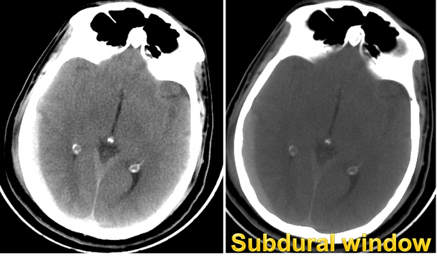
CT head (subdural window)
Citation, DOI, disclosures and article data
Citation:
Knipe H, Murphy A, Toumpanakis D, et al. CT head (subdural window). Reference article, Radiopaedia.org (Accessed on 27 Mar 2025) https://doi.org/10.53347/rID-48361
rID:
48361
Article created:
Disclosures:
At the time the article was created Henry Knipe had no recorded disclosures.
View Henry Knipe's current disclosures
Last revised:
Disclosures:
At the time the article was last revised Andrew Murphy had no financial relationships to ineligible companies to disclose.
View Andrew Murphy's current disclosures
Revisions:
9 times, by
8 contributors -
see full revision history and disclosures
Systems:
Sections:
Synonyms:
- Blood window
- Subdural window
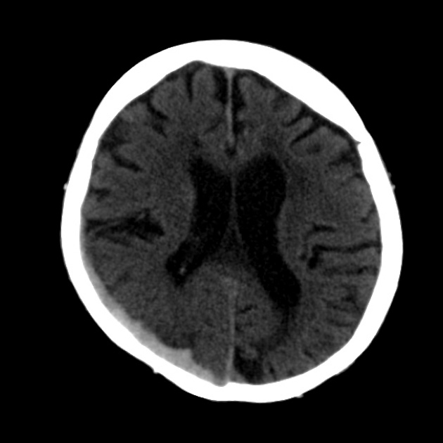
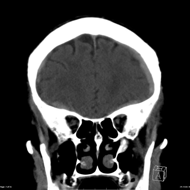
The subdural (blood) window can be used when reviewing a CT brain as it makes intracranial haemorrhage more conspicuous, and may help in the detection of thin acute subdural haematomas that are against the calvaria that are similar density to adjacent bone. It is a wider setting than the standard non-contrast window, and there are a number of parameters that can be used:
- centre/level 50 HU; width 130 HU 1
- centre/level 70-100 HU; width 150-300 HU 3
Quiz questions
References
- 1. Emergency Radiology (Rotations in Radiology). Oxford University Press. ISBN:0190223650. Read it at Google Books - Find it at Amazon
- 2. Brant WE, Helms CA. Fundamentals of Diagnostic Radiology. Lippincott Williams & Wilkins. (2007) ISBN:0781761352. Read it at Google Books - Find it at Amazon
- 3. Diseases of the Brain, Head & Neck, Spine. Springer. ISBN:8847008395. Read it at Google Books - Find it at Amazon
Incoming Links
Articles:
Cases:
Multiple choice questions:
Related articles: Computed tomography
- computed tomography in practice
-
computed tomography overview
- iodinated contrast media
- CT IV contrast media administration
-
CT artifacts
- patient-based artifacts
- physics-based artifacts
- hardware-based artifacts
- ring artifact
- tube arcing
- out of field artifact
- air bubble artifact
- helical and multichannel artifacts
- CT technology
-
generations of CT scanners
- helical CT scanning
- step and shoot scanning
- ultra-high-resolution CT (UHRCT)
- CT x-ray tube
- CT fluoroscopy
- cone-beam CT
-
generations of CT scanners
- dual-energy CT
- CT image reconstruction
- CT image quality
- CT dose
-
CT protocols
- composite
- head & neck
- chest
- abdomen and pelvis
- CT abdomen-pelvis (protocol)
- CT abdominal aorta
- CT adrenals (protocol)
- CT cholangiography (protocol)
- CT colonography (protocol)
- CT enteroclysis (protocol)
- CT enterography (protocol)
- CT gastrography (protocol)
- CT kidneys, ureters and bladder (protocol)
- CT urography (protocol)
- CT Renal mass (protocol)
- CT angiography of the splanchnic vessels (protocol)
- CT renal split bolus
- CT pancreas (protocol)
- liver


 Unable to process the form. Check for errors and try again.