
Elbow ossification
Citation, DOI, disclosures and article data
At the time the article was created Frank Gaillard had no recorded disclosures.
View Frank Gaillard's current disclosuresAt the time the article was last revised Tariq Walizai had no financial relationships to ineligible companies to disclose.
View Tariq Walizai's current disclosures- Ossification of elbow
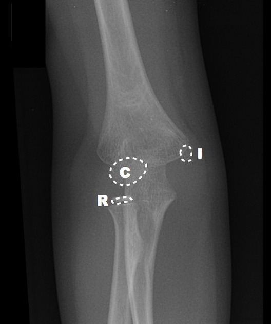
Elbow ossification occurs at the six elbow ossification centres in a reproducible order. Being familiar with the order of ossification of the elbow is important in not mistaking an epicondylar fracture for a normal ossification centre.
Appearance
Order
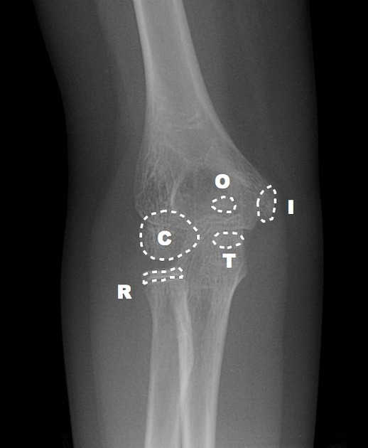
The order of appearances of the elbow ossification centres is highly reliable and in most individuals, is consistent: capitellum, radial head, internal (medial) epicondyle, trochlea, olecranon and external (lateral) epicondyle.
The order of "I" and "T" are most important to remember; the trochlea ossification centre should not appear before the internal (medial) epicondyle ossification centre. If you can see a trochlea but no internal epicondyle, then you need to look very hard for the avulsed ossification centre.
Age
Two counting methods are taught to help remember the ages at which the ossification centres appear: 1-3-5-7-9-11 (simple) and 1-5-7-10-10-11 (more accurate).
capitellum: 1 year
radial head: 3 years
internal epicondyle: 5 years
trochlea: 7 years
olecranon: 9 years
external epicondyle: 11 years
Therefore, if the trochlear centre is present, but there is no medial epicondyle then you are most likely looking at a medial epicondylar fracture where the ossification centre has been avulsed and displaced. This is extremely important since these injuries should be seen by an orthopaedic surgeon and internally fixed.
Variation
Exceptions have been described in the literature to the usual sequence of appearance of elbow ossification centres and recognised as normal variants 2. This can occur in up to a quarter of children with incidence much more common in girls.
Video
References
- 1. Iyer RS, Thapa MM, Khanna PC et-al. Pediatric bone imaging: imaging elbow trauma in children-a review of acute and chronic injuries. AJR Am J Roentgenol. 2012;198 (5): 1053-68. doi:10.2214/AJR.10.7314 - Pubmed citation
- 2. SJ Goodwin, LJ Irwin, GJ Irwin. Gender differences in the order of appearance of elbow ossification centres:. (2018) Scottish Medical Journal. 64 (1): 2-9. doi:10.1177/0036933018812000 - Pubmed
Incoming Links
- Paediatric elbow anatomy (illustrations)
- Un-united medial epicondyle ossification centre
- Lateral condyle fracture
- Non-united medial epicondyle ossification center
- Medial epicondyle fracture
- Elbow radiograph - age eleven
- Elbow radiograph - age nine
- Elbow radiograph - age five
- Elbow radiograph - age four
- Elbow radiograph - age two
Related articles: Anatomy: Upper limb
-
skeleton of the upper limb
- clavicle
- scapula
- humerus
- radius
- ulna
- hand
- accessory ossicles of the upper limb
- accessory ossicles of the shoulder
- accessory ossicles of the elbow
-
accessory ossicles of the wrist (mnemonic)
- os centrale carpi
- os epilunate
- os epitriquetrum
- os styloideum
- os hamuli proprium
- lunula
- os triangulare
- trapezium secondarium
- os paratrapezium
- os radiostyloideum (persistent radial styloid)
- joints of the upper limb
-
pectoral girdle
-
shoulder joint
- articulations
- associated structures
- joint capsule
- bursae
- ligaments
- movements
- scapulothoracic joint
-
glenohumeral joint
- arm flexion
- arm extension
- arm abduction
- arm adduction
- arm internal rotation (medial rotation)
- arm external rotation (lateral rotation)
- circumduction
- arterial supply - scapular anastomosis
- ossification centres
-
shoulder joint
-
elbow joint
- proximal radioulnar joint
- ligaments
- associated structures
- movements
- alignment
- arterial supply - elbow anastomosis
- development
-
wrist joint
- articulations
-
ligaments
- intrinsic ligaments
- extrinsic ligaments
- radioscaphoid ligament
- dorsal intercarpal ligament
- dorsal radiotriquetral ligament
- dorsal radioulnar ligament
- volar radioulnar ligament
- radioscaphocapitate ligament
- long radiolunate ligament
- Vickers ligament
- short radiolunate ligament
- ulnolunate ligament
- ulnotriquetral ligament
- ulnocapitate ligament
- ulnar collateral ligament
- associated structures
- extensor retinaculum
- flexor retinaculum
- joint capsule
- movements
- alignment
- ossification centres
-
hand joints
- articulations
- carpometacarpal joint
-
metacarpophalangeal joints
- palmar ligament (plate)
- collateral ligament
-
interphalangeal joints
- palmar ligament (plate)
- collateral ligament
- movements
- ossification centres
- articulations
-
pectoral girdle
- spaces of the upper limb
- muscles of the upper limb
- shoulder girdle
- anterior compartment of the arm
- posterior compartment of the arm
-
anterior compartment of the forearm
- superficial
- intermediate
- deep
-
posterior compartment of the forearm (extensors)
- superficial
- deep
- muscles of the hand
-
accessory muscles
- elbow
- volar wrist midline
- palmaris longus profundus
- aberrant palmaris longus
- volar wrist radial-side
- accessory flexor digitorum superficialis indicis
- flexor indicis profundus
- flexor carpi radialis vel profundus
- accessory head of the flexor pollicis longus (Gantzer muscle, common)
- volar wrist ulnar-side
- dorsal wrist
- blood supply to the upper limb
-
arteries
- subclavian artery (mnemonic)
- axillary artery
- brachial artery (proximal portion)
- ulnar artery
- radial artery
- veins
-
arteries
- innervation of the upper limb
- intercostobrachial nerve
-
brachial plexus (mnemonic)
- branches from the roots
- branches from the trunks
- branches from the cords
- lateral cord
- posterior cord
- medial cord
- terminal branches
- lymphatic drainage of the upper limb


 Unable to process the form. Check for errors and try again.