
Fetal ovarian cysts refer to an ovarian cyst detected antenatally in a female fetus. They are relatively uncommon and are usually diagnosed in the 3rd trimester 5.
On this page:
Epidemiology
From autopsy studies, ovarian cysts are found in up to 30% of fetuses 1.
Associations
Other associated anomalies are considered generally rare 2.
Pathology
The exact aetiology of fetal ovarian cysts is not well known but it is thought to be related to some form of hormonal stimulation (e.g. fetal gonadotropins, maternal oestrogen, and placental beta-HCG) 6.
A fetal ovarian cyst can be of variable size. It is not thought to change significantly in size over the latter course of pregnancy 2.
Location/distribution
They tend to be unilateral although bilateral cysts are also rarely seen.
Radiographic features
Ultrasound
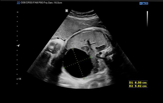
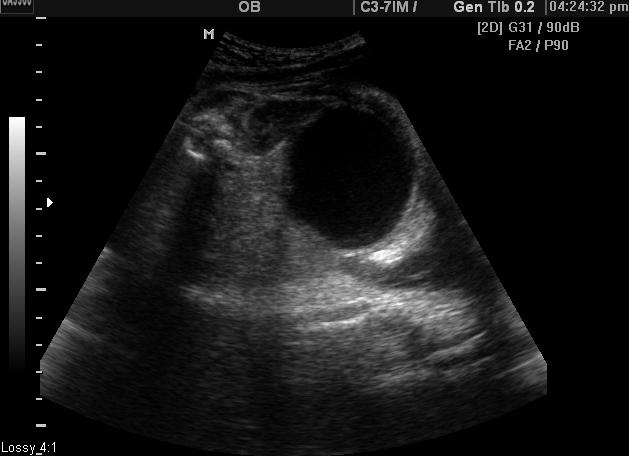
While it is often difficult to accurately diagnose a fetal ovarian cyst sonographically due to many other cystic lesions having similar appearances, it is typically seen as a well-circumscribed non-septated cyst in the fetal pelvis separate from the fetal bladder, stomach and gallbladder. It is often anechoic if simple and uncomplicated. The presence of a small daughter cyst is considered a characteristic feature. Due to the relative laxity of supporting ligaments, the cyst can sometimes rise into the upper abdomen.
If there has a complication there may be fetal ascites (from cyst rupture) or irregular echogenic intracystic material (from a haemorrhage).
Complications
in utero cyst rupture
cyst torsion
intracystic haemorrhage
compression of adjacent structures
Treatment and prognosis
They are almost always benign simple cysts. Treatment is usually conservative and in selected cases antenatal or neonatal cyst aspiration, laparoscopic cystectomy and laparotomy have been considered. Some advocate intervention if cysts are complex, wanders about the abdomen on serial scans, are larger than 4-5 cm or demonstrate rapid interval enlargement 4,5,7. Spontaneous remission can commonly occur although on occasion can take up several months 11.
Differential diagnosis
Differential considerations for a cystic lesion in the fetal pelvis include:
non-calcified meconium pseudocyst
hydrocolpos 12


 Unable to process the form. Check for errors and try again.