
Focal hepatic steatosis, also known as focal hepatosteatosis or (erroneously) focal fatty infiltration, represents small areas of liver steatosis. In many cases, the phenomenon is believed to be related to the hemodynamics of a third inflow.
On this page:
Epidemiology
Essentially the same as those that contribute to diffuse hepatic steatosis 1,5:
alcohol use
exogenous steroids
drugs (amiodarone, methotrexate, chemotherapy)
IV hyperalimentation
In general, the treatment of the underlying condition will reverse the findings.
Pathology
Location
A characteristic location for focal hepatosteatosis is the medial segment of the left lobe of the liver (segment 4) either anterior to the porta hepatis or adjacent to the falciform ligament 1. This distribution is the same as that seen in focal fatty sparing and is thought to relate to variations in vascular supply. This also would account for focal fatty change/sparing sometimes seen related to vascular lesions.
Radiographic features
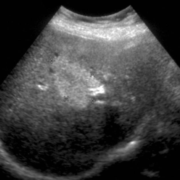
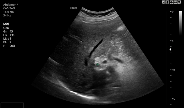
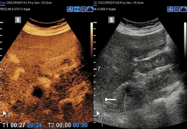
Ultrasound
Ultrasound features only become apparent when the amount of fat reaches 15-20%. Features include:
hyperattenuation of the beam
mild or absent positive mass effect
geographic borders
no distortion of vessels
inability to visualize the portal vein walls (as the parenchyma is as bright as the wall)
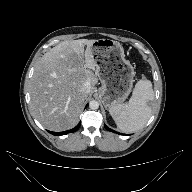
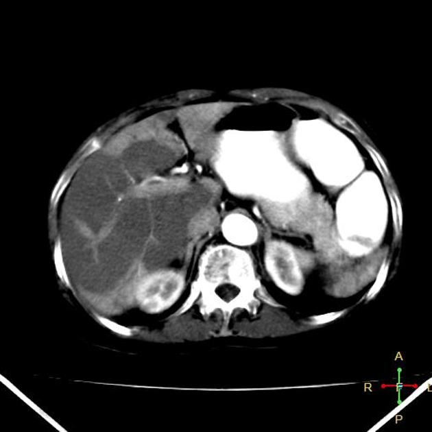
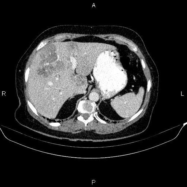
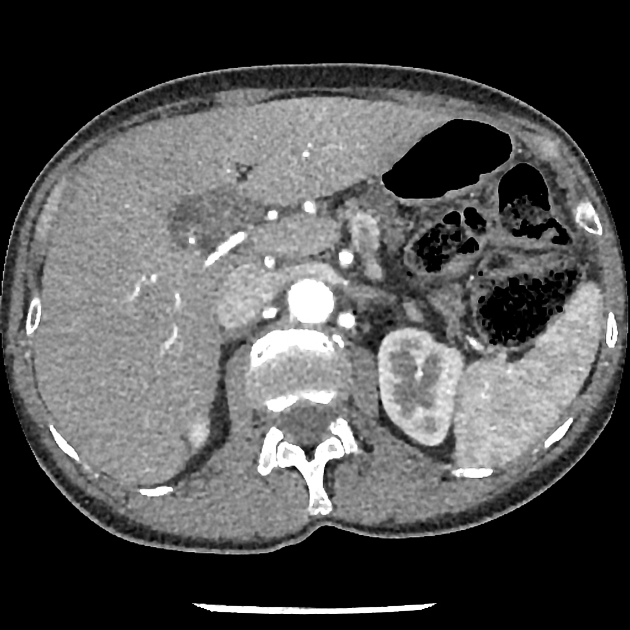
CT
-
decreased attenuation (non-contrast CT)
normal liver 50-57 HU
decreases by 1.6 HU per mg of fat in each gram of liver
-
decreased attenuation (post-contrast CT)
liver and spleen should normally be similar on delayed (70 seconds) scans
earlier scans are unreliable as the spleen enhances earlier than the liver (systemic supply rather than portal)
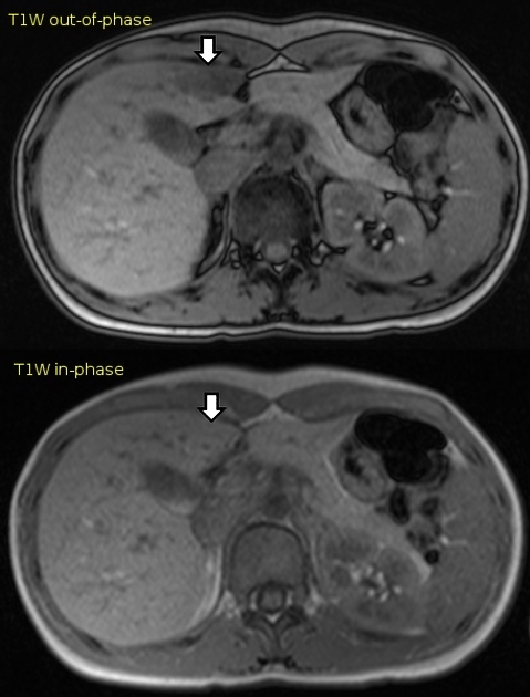
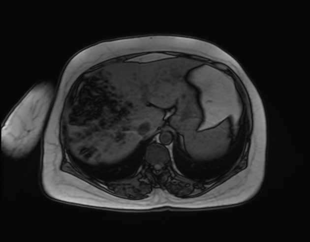
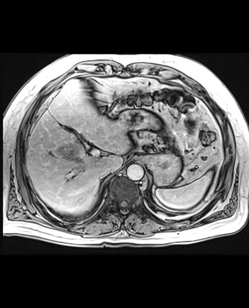
MRI
MRI is the imaging modality of choice in any case where the diagnosis is felt to be less than certain
increased T1 signal
signal drop-out on opposed-phase imaging
ability to quantify the fat fraction
Differential diagnosis
When located in characteristic locations then there is usually little difficulty in making the correct diagnosis. If unusual in location or appearance then differentials to be considered include:
-
the commonest hyperechoic liver lesion, typically well defined and may show peripheral feeding vessels


 Unable to process the form. Check for errors and try again.