
Presentation
Abdominal pain.
Patient Data


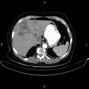

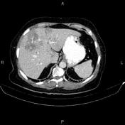
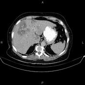
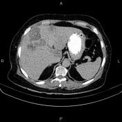
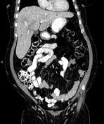
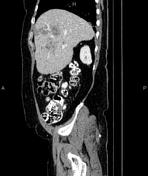
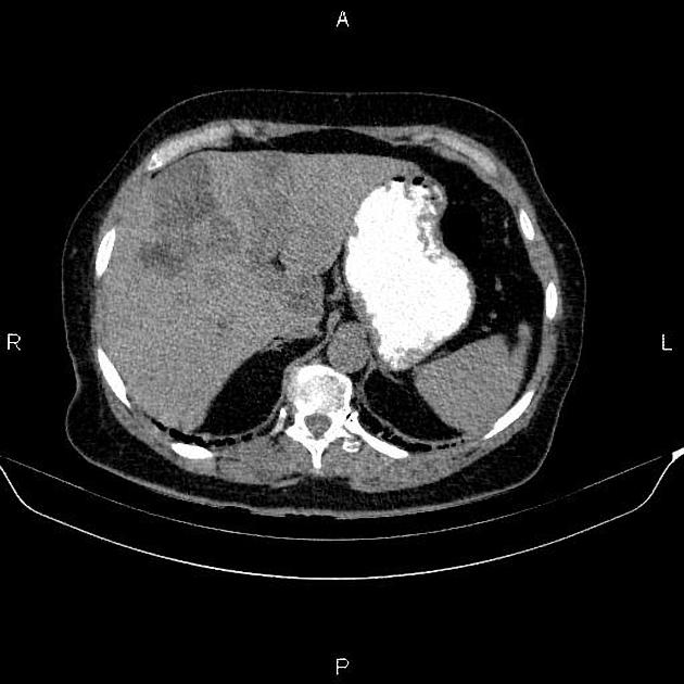
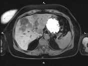
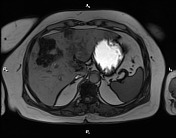
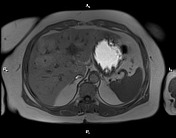
A lobulated geographic hypodense area is seen occupying most of the right lobe and the medial segment of the left lobe with the blood vessels seen transversing this region without interruption. The lesion becomes less distinct in delayed images. No mass effect could be detected.
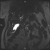
Mild intrahepatic bile duct dilatation is seen.

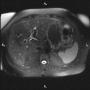


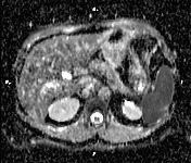
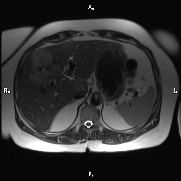
Signal dropout on opposed phase T1W GRE images is noted in the 4th and 8th hepatic segments in favor of fatty infiltration.
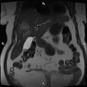
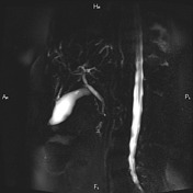
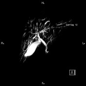
Alternating dilated and stenotic segments of the intrahepatic bile ducts are visible.
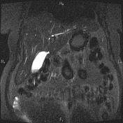
Mild irregularity of the common bile duct. Low signal filling defect is noted in the gallbladder.
Subtle periportal edema is present.
Case Discussion
Biliary duct changes related to primary sclerosing cholangitis (known case) which is an uncommon inflammatory condition and affects the biliary tree resulting in multiple strictures, liver damage, and eventually cirrhosis.
Patchy focal fat deposition and relative fatty sparing may be mistaken for a focal neoplasm (particular on US and CT) and thus MR in-phase and opposed-phase imaging allows reliable differentiation


 Unable to process the form. Check for errors and try again.