
Greater tubercle fractures of the shoulder are a subtype of proximal humeral fractures.
On this page:
Gross anatomy
The greater tubercle is the most lateral bony part of the shoulder. It is the site where three of the rotator cuffs insert to abduct or laterally rotate the shoulder joint (supraspinatus, infraspinatus, and teres minor). In the setting of fractures, the greater tubercle fragment can be pulled superiorly by supraspinatus or posteriorly by teres minor.
arterial supply: posterior humeral circumflex artery and the arcuate artery (branch of the anterior circumflex)
Epidemiology
Greater tubercle fractures are more common in the setting of high velocity trauma, i.e. skiing or motor vehicle accidents, or in combination with another proximal humeral fracture. Despite this, isolated greater tubercle fractures only account for 2-19% of proximal humeral fractures 1.
Nevertheless, the majority of greater tubercle fractures are minimally displaced and are treated conservatively.
Clinical presentation
Clinical features include:
pain on rest and on palpation
swelling
decreased motion
Pathology
Greater tubercle fractures can be divided into:
avulsion fractures
depression fractures
split fractures
Radiographic features
Plain radiograph
Greater tubercle fractures can be subtle and subsequently missed on initial radiographic imaging. Improved detection may occur with apical oblique projections and AP projections with external rotation.
X-ray typically shows incongruence of the bone on external AP and axillary views.
Ultrasound
Ultrasound is helpful in diagnosing fractures where there has been concurrent rotator cuff tears.
Ultrasound features may include discontinuity, sharp defect, or irregularity of cortical bone in the greater tubercle. However, such findings are non-specific, as advanced impingement can give this appearance 2
CT
If a fracture is not detected on initial radiographic imaging, CT may be obtained if there is a high index of suspicion.
There is ongoing debate whether CT provides any further benefit compared to plain radiography alone. Literature suggests that CT does not lead to a change in treatment recommendation compared to plain radiographs. Nevertheless, it does increase surgeons' confidence for recommendation of treatment 3.
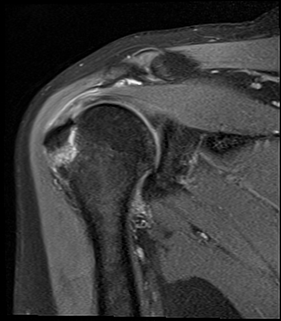
MRI
MRI can be performed where there is clinical suggestion of a concomitant rotator cuff tear. MRI is relatively sensitive in diagnosing greater tubercle fractures. Greater tubercle fractures are best diagnosed on oblique coronal T1 and T2 weighted images 3,4.
MRI features include:
oblique coronal T1 and T2: shows crescentic or oblique lines of decreased signal intensity extending to the cortical bone
STIR and T1: shows adjacent marrow edema in the affected medullary bone
Treatment and prognosis
Proximal humeral fractures are classified according to the Neer classification. However, there is an absence of criteria specific to isolated greater tubercle fractures 5. Management includes:
non-operative: minimally displaced fractures can be treated conservatively with immobilization and rehabilitation
-
surgical: displaced postero-superior fractures >5 mm can be considered with reduction and internal fixation
there is controversy in the literature regarding the degree of displacement requiring fixation; nevertheless, the majority of the literature agrees that >5 mm of displacement has a greater risk of malunion
Differential diagnosis
on plain radiograph, enthesophytosis can sometimes mimic a greater tubercle fracture


 Unable to process the form. Check for errors and try again.