
Hoffa fat pad ganglion cysts are intra-articular ganglion cysts and are the most common mass-like lesions within the Hoffa fat pad.
On this page:
Epidemiology
Hoffa fat pad ganglion cysts are rare and less common than cruciate ligament ganglion cysts 1,2.
Clinical presentation
Ganglia within Hoffa’s fat pad can be asymptomatic or can cause pain and swelling or present as a palpable mass 1,2.
Pathology
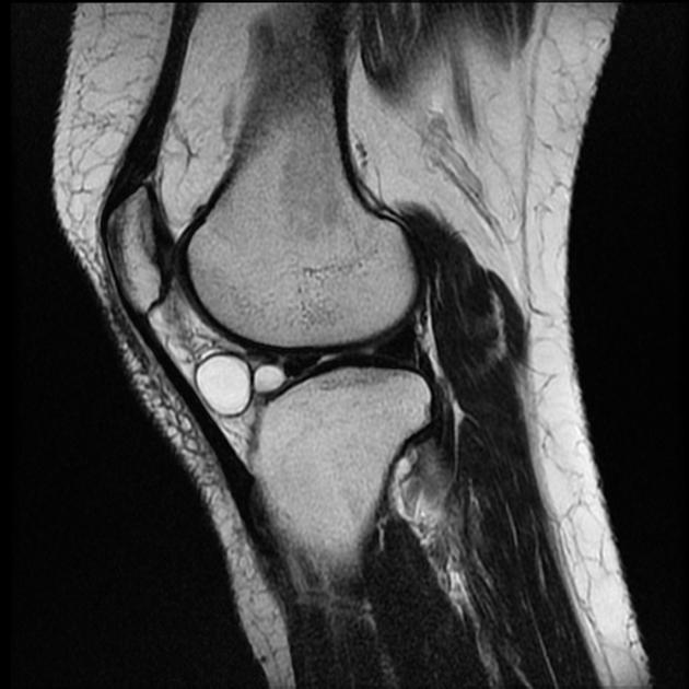
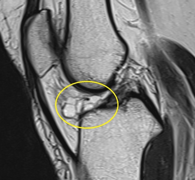
Ganglion cysts within Hoffa’s fat pad are usually well-defined, smooth-walled, uni- or multilocular, lobulated cystic masses surrounded by dense connective tissue and filled with viscous, mucinous material like other ganglion cysts. They lack a synovial membrane 1-3.
Aetiology
Like with other ganglion cysts, the pathogenesis is controversial and comprises the following theories 2-4:
a sequel of mucoid degeneration
cyst formation as a consequence of trauma or tissue irritation
release of hyaluronic acid by mesenchymal stem cells and consecutive cyst formation
synovial herniation
congenital translocation of synovial cells
Location
Ganglion cysts of Hoffa’s fat pad are usually located anterior to the anterior horn of the lateral meniscus 1.
Radiographic features
MRI
MRI is the best modality for the visualisation and evaluation of intra-articular ganglion cysts. The typical appearance is that of a well-defined lobular cystic mass within or adjacent to Hoffa’s fat pad 1,2,4.
T1: usually hypointense but will depend on protein content
T2: hyperintense
PDFS/T2FS: hyperintense
Treatment and prognosis
Management options depend vastly on clinical symptoms and include conservative measures, image-guided percutaneous aspiration as well as arthroscopic or surgical excision 2,4-6.
Differential diagnosis
The differential is that of cyst-like lesions around the knee:


 Unable to process the form. Check for errors and try again.